

Bedside Snapshot
- Recognize
- Muscle injury with CK above 5 times the upper limit of normal or above 1,000 U/L, often with dark urine and weakness. Crush or prolonged entrapment. Compartment syndrome: pain out of proportion, pain on passive stretch, tense limb.
- Red Flags
- Potassium above 6 mEq/L or any hyperkalemic ECG change; urine output under 1 mL/kg/hr; tense compartment with pain on stretch; absent distal pulse; confusion or shock
- First Actions
- Monitor and 12-lead ECG; start IV crystalloid early and titrate to urine output; calcium for any hyperkalemic ECG change; insulin with dextrose; serial potassium and CK; surgical consult for suspected compartment syndrome
- Key Doses
- 0.9% saline or lactated Ringer's about 400 mL/hr to start, titrated to urine output 1 to 3 mL/kg/hr
- Calcium gluconate 10% 3 g IV over 10 min for ECG change
- Regular insulin 10 units IV with dextrose 25 g
- Don't Miss
- Compartment syndrome with a palpable pulse or a normal-looking limb. Hyperkalemia that appears after release. Pain out of proportion comes first; pulselessness is late.
- Escalate & Transport
- Urgent surgical consult for suspected compartment syndrome; nephrology for dialysis if AKI or refractory hyperkalemia. ICU or monitored unit for arrhythmia, shock or rising potassium. Transport to a center with surgery, dialysis and ICU.
Definition & Pathophysiology
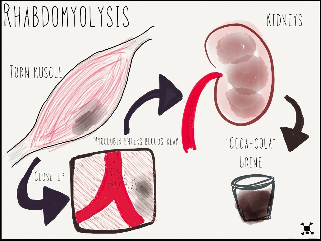
Rhabdomyolysis is muscle cells dying and spilling their contents into the blood: creatine kinase (CK), myoglobin, potassium, phosphate and lactate dehydrogenase. The AAST 2022 consensus holds no formal definition. The lab cutoff most often used is CK above 5 times the upper limit of normal or above 1,000 U/L, with weakness, pain or swelling. Crush syndrome is the systemic form. It follows prolonged compression of tissue, and reperfusion adds a second injury.
Compartment syndrome is pressure rising inside a closed fascial compartment. Swelling or bleeding in the fascia squeezes the capillaries. Ischemia follows, then more swelling and more pressure. Muscle damage can become irreversible if the diagnosis waits more than 6 hours.
- Traumatic causes: Crush injury, entrapment, prolonged immobility (found down), fractures, electrical and burn injury, and vascular injury. Highest risk: multisystem trauma, extremity or torso crush, and compartment syndrome.
- Non-traumatic causes: Strenuous exercise, seizures, alcohol, cocaine and amphetamines, viral infection, temperature extremes and inherited metabolic myopathies. Drugs on the list include statins, propofol, daptomycin and colchicine.
- Clinical course: CK peaks 24 to 72 hours after the insult. Acute kidney injury (AKI) is the most common serious complication, alongside hyperkalemia, arrhythmia, DIC and compartment syndrome. The AAST 2022 consensus puts mortality among inpatients with CK above 5,000 U/L at about 14%. Acute renal failure develops in up to 15% of patients. Rates vary by population and definition.
Recognition & Red Flags
Presentations
- Rhabdomyolysis: Muscle pain, tenderness and weakness, swelling of the involved muscle group, and dark (cola-colored) urine. Some patients have none of this. Only a high CK gives it away.
- Crush injury: Bruising, swelling and pain over the compressed limb or torso. Blisters or pressure ulcers on the skin may suggest it. Don't bank on them, because they are not reliable.
- Compartment syndrome: Pain out of proportion to the injury. That is the earliest sign. Pain on passive stretch of the muscles in that compartment, a tense, firm limb, and numbness or paresthesia. Pallor, pulselessness and paralysis are the late signs.
Red Flags
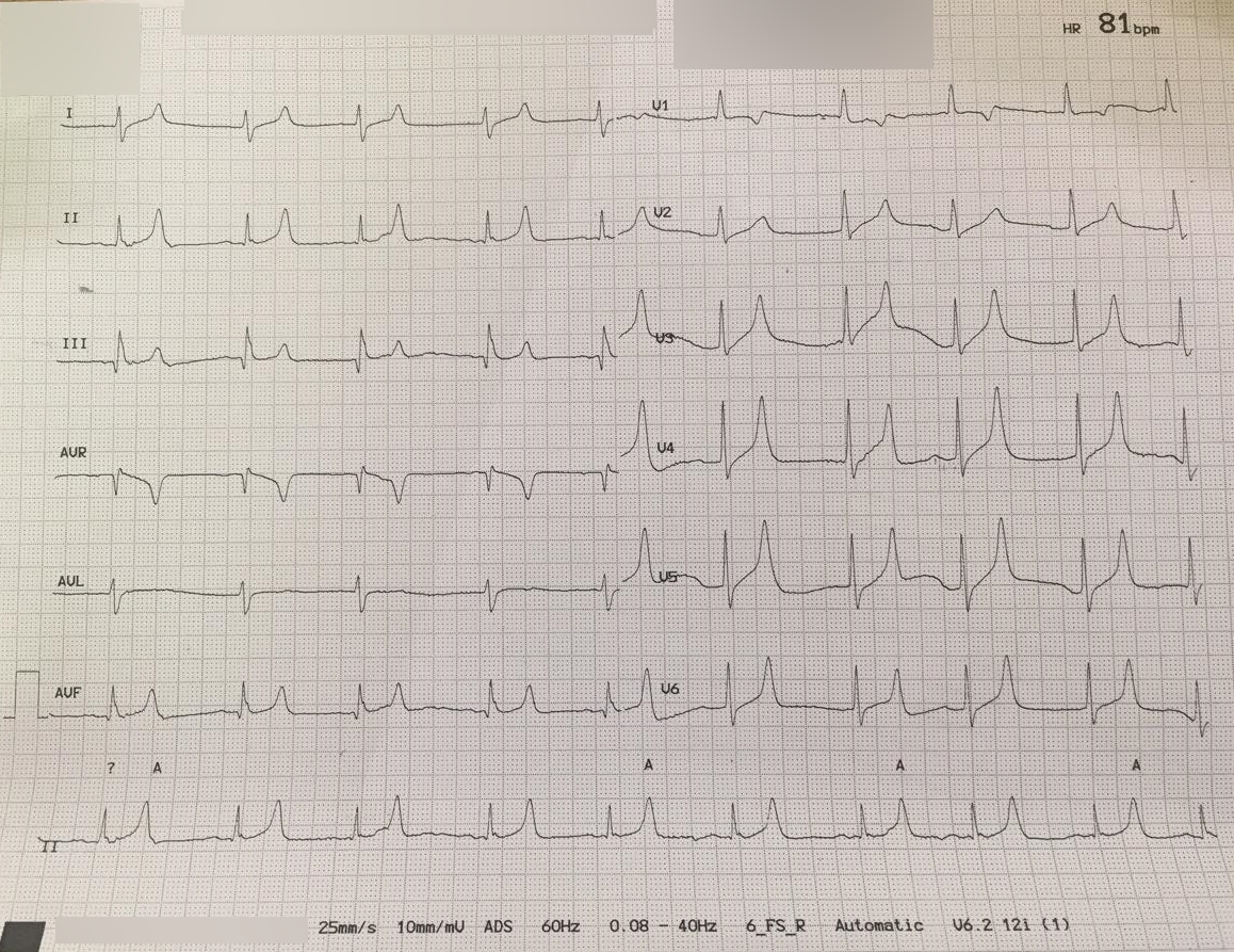
- Potassium above 6 mEq/L, or any hyperkalemic ECG change (peaked T waves, PR or P-wave loss, QRS widening, bradycardia)
- Urine output under 1 mL/kg/hr after fluid is started, or dark urine with no red cells on microscopy
- Pain on passive stretch, a tense compartment, or new numbness: suspect compartment syndrome now
- Absent distal pulse in an injured limb: emergent surgical evaluation for compartment syndrome or vascular injury
- Confusion, hypotension, shock or new arrhythmia
Assessment & Diagnostics
Three labs anchor the workup: potassium, CK and kidney function. Where you can get it, compartment pressure measurement is the most reliable test for compartment syndrome.
| Check | Why | Act If |
|---|---|---|
| 12-lead ECG and rhythm | Hyperkalemia changes conduction before the potassium result returns. See electrolyte ECG | Any hyperkalemic change: calcium now and continuous monitoring (ALS section) |
| Potassium (K⁺) | Rises early and tracks the amount of muscle destroyed. See potassium | Above 6 mEq/L: continuous monitoring and serial levels, per the AAST 2022 consensus. Treat per the Hyperkalemia page |
| CK, serial | Confirms rhabdomyolysis at 5 times the upper limit of normal or above 1,000 U/L. Peaks at 24 to 72 hours; above 5,000 U/L carries higher AKI risk | Repeat until the peak is passed and falling. Many sources continue IV fluid until CK falls below 5,000 U/L |
| Urinalysis | Dipstick positive for blood with no red cells on microscopy suggests myoglobinuria. Urine myoglobin is up to 100% sensitive but only 15% to 88% specific in the AAST 2022 consensus, so the dipstick cannot separate myoglobin from hemoglobin. See urine blood | Positive with a raised CK: rhabdomyolysis, so start fluids. Serum myoglobin, see myoglobin, has poor sensitivity for rhabdomyolysis, so follow CK |
| Creatinine, calcium, phosphate, bicarbonate | Creatinine rise means AKI. Low calcium, high phosphate and low bicarbonate are McMahon score items. See creatinine and calcium | Rising creatinine or oliguria: AKI pathway and nephrology. Low calcium alone: record it and treat only if symptomatic or ECG changes are present |
| McMahon score | Calculated at admission from age, sex, creatinine, CK, calcium, phosphate, bicarbonate and cause. A score of 6 or more predicts the need for high-volume fluid, RRT or death. AAST 2022 reports 86% sensitivity and 68% specificity for RRT | 6 or more: high-volume fluid resuscitation per AAST 2022, with early ICU and nephrology input per local protocol |
| Compartment pressure | Measured with a pressure monitor where available. Adult thresholds: absolute pressure above 30 mmHg, or delta pressure (diastolic BP minus compartment pressure) under 30 mmHg | Threshold met, or strong clinical suspicion: urgent surgical consult for fasciotomy |
| Blood glucose | Required before insulin. Hypoglycemia is the main risk of insulin for hyperkalemia | Under 126 mg/dL before insulin: add the glucose infusion in the ALS section |
Don't trust the exam alone. In a meta-analysis of tibial injuries, the predictive value was 21% for clinical signs and 29% for pressure measurement. Combined, the two reached 68%.
Differential & Mimics
- Statin-associated myopathy: Diffuse aches and a raised CK. No myoglobinuria, no AKI and no swollen compartment.
- Autoimmune myositis: Subacute weakness, rash, Raynaud phenomenon or interstitial lung symptoms. Still no myoglobinuria or kidney injury.
- Liver injury: AST and ALT rise with muscle injury too. In acute rhabdomyolysis the AST:ALT ratio can go above 3, and both enzymes fall along with CK. Suspect liver disease only when they do not fall together.
- Hematuria or hemoglobinuria: Red cells on microscopy point to bleeding. The dipstick cannot tell hemoglobin from myoglobin, so a positive dipstick with no red cells still needs a CK.
BLS Management
- Scene safety: do not enter unstable debris. Keep the patient still and protect the crushed limb.
- Record the time: when the limb was compressed and when it was released. Longer entrapment predicts acute renal failure.
- Monitor: pulse, blood pressure, SpO₂, mental status and urine color. Ask about urine output and any dark urine.
- Support: oxygen for hypoxia and a position that eases breathing. Keep the limb level and still.
- Check the limb and report: pain, tenseness of the compartment, numbness and pulses. Call ALS early and report the time of entrapment, the mechanism, any medicines and the suspicion of compartment syndrome.
ALS Management
Doses vary by protocol; follow local protocols and medical direction. Start fluid early and titrate it to urine output. For any ECG change, calcium goes first. The potassium shift and removal come after.
- Monitoring (Paramedic/RN/CCT): Continuous cardiac monitoring and a 12-lead ECG when the protocol allows. Keep the monitor on for potassium above 6 mEq/L, per the AAST 2022 consensus. Keep it on for any ECG change too, and for an unwell patient with a potassium of 6.0 to 6.4 mEq/L.
- IV or IO access (Paramedic/RN/CCT): large-bore access. Use IO access if IV fails.
- Crystalloid (Paramedic/RN/CCT): Start 0.9% saline or lactated Ringer's at about 400 mL/hr, before extrication where protocol allows. AAST 2022 calls a starting range of 200 to 1,000 mL/hr reasonable. Titrate to a urine output of 1 to 3 mL/kg/hr. The AAST figure of up to about 300 mL/hr marks the upper urine-output target. It is not a cap on the IV rate. Stop boluses for crackles, falling SpO₂ or rising work of breathing. Protocols differ on fluid type. 0.9% saline has no potassium. The lactated Ringer's label advises avoiding it, or monitoring potassium closely, in tissue injury. See Normal Saline and Lactated Ringer's
- Calcium for any hyperkalemic ECG change (Paramedic/RN/CCT): calcium gluconate 10% 3 g IV over 10 min per the 2026 UKKA guideline (label rate limit 200 mg/min), or calcium chloride 10% 1 g IV slowly, into a patent large vein. That 3 g gluconate dose comes from the guideline, not the label. The label's adult bolus range for hypocalcemia is 1 to 2 g, so follow protocol for the dose. Repeat if the ECG change persists. Don't give calcium for asymptomatic hypocalcemia in rhabdomyolysis. AAST 2022 advises avoiding early treatment unless the patient is symptomatic or has severe hyperkalemia, because calcium can deposit in injured muscle and rebound to hypercalcemia in recovery. Treat low calcium when there is an ECG change or symptoms such as tetany, seizures or arrhythmia. See Calcium Gluconate
- Insulin with dextrose (Paramedic/RN/CCT): regular insulin 10 units IV with dextrose 25 g for potassium 6.5 mEq/L or above, or an ECG change, per the Hyperkalemia page. Hypoglycemia risk runs higher with acute kidney injury or CKD, low body weight and no diabetes. UKKA 2026 keeps 10 units. Lower regimens reduce hypoglycemia risk, so follow local protocol for a reduced dose. See Insulin and Dextrose
- Glucose check (Paramedic/RN/CCT): Check glucose before insulin, then at 30, 60, 90, 120, 180, 240, 300 and 360 minutes after it (UKKA 17.2, as on the Hyperkalemia page). Hypoglycemia can occur up to 6 hours after the dose, which is why the checks run this long. If glucose was under 126 mg/dL before treatment, give 10% glucose at 50 mL/hr for 5 hours (25 g), per the Hyperkalemia page.
- Albuterol, adjunct only (Paramedic/RN/CCT): nebulized albuterol 10 to 20 mg for potassium 6.5 mEq/L or above, optional at 6.0 to 6.4 (UKKA 2026, as on the Hyperkalemia page). It is an adjunct and never the only treatment for hyperkalemia. In ischemic heart disease, use 10 mg. See Albuterol
- Do not give routinely (Paramedic/RN/CCT): sodium bicarbonate or mannitol to prevent AKI. The AAST 2022 consensus does not recommend either for this purpose. UKKA 2026 does not make bicarbonate routine for hyperkalemia. Give it for systemic metabolic acidosis per protocol, on a separate line from calcium. See Sodium Bicarbonate
- Analgesia (Paramedic/RN/CCT): Give pain relief as needed. Record the pain score and the analgesic need before and after each dose. A rising need for analgesia can be the first sign of compartment syndrome.
ED & ICU Management
- Labs and serial checks: Potassium, CK until its peak, creatinine, calcium, phosphate, bicarbonate, glucose and coagulation studies. DIC often runs silently. Expect prolonged clotting times, low platelets and raised fibrin degradation products, with no bleeding to show for it.
- Fluid: Keep IV crystalloid running for at least the first 24 hours, per Usuda 2023. Titrate to urine output of 1 to 3 mL/kg/hr, per AAST 2022. Many sources keep fluid going until CK falls below 5,000 U/L. Weigh that against volume overload, which is more likely in older adults and in patients with heart or kidney disease.
- Hyperkalemia: Run it per the Hyperkalemia page. Calcium for any ECG change, insulin with dextrose, potassium removal, and dialysis for refractory hyperkalemia. Expect rebound after the insulin shift.
- Nephrotoxins: Stop nephrotoxins that are making the injury worse. Check for causative drugs, including statins, propofol, daptomycin and colchicine, and involve the prescriber.
- Kidney replacement: Start RRT for the standard AKI indications. In rhabdomyolysis with AKI, choose CRRT or intermittent RRT based on the degree of impairment and clinical status. RRT has no role in preventing AKI. Dialysis clears myoglobin poorly. See Hemodialysis
- Compartment syndrome: Urgent surgical consult. Emergent, wide-incision decompressive fasciotomy is the standard for established compartment syndrome. Fasciotomy in crush injury has not been shown to improve outcome. Delayed fasciotomy is linked to worse function, though, so keep the threshold low. Fasciotomy carries bleeding and wound-sepsis risk.
- Consults and disposition: Call nephrology for AKI or refractory hyperkalemia. Surgery handles compartment decompression. ICU takes arrhythmia, shock, or any airway or breathing problem.
Transport Considerations
- Destination: Go to a center with surgical capability for compartment decompression, dialysis and an ICU. Protocol decides which center. Don't let the transfer delay the surgical decision.
- Continue and monitor: Keep the monitor on. Repeat the ECG after calcium. Keep the IV fluid on its pump, track hourly urine output, and check the compartments and pulses at each reassessment.
- Hyperkalemia risk: Potassium can climb shortly after extrication or reperfusion. Risk peaks right after extrication, so keep monitoring after you leave the scene.
- Handoff essentials: Hand off the time of entrapment and release, fluid volumes with times, potassium and CK values with times, ECG before and after treatment, each calcium, insulin and dextrose dose with its time, urine output, the glucose series, and all medicines.
- Equipment: calcium gluconate and calcium chloride, regular insulin, 50% dextrose, 10% dextrose for the glucose infusion where protocol carries it, nebulized albuterol, 0.9% saline and lactated Ringer's, a glucometer, a monitor that records a 12-lead, and a compartment pressure monitor where carried.
Special Populations
- Pediatrics: Fluid is a 10 to 20 mL/kg IV/IO bolus over 5 to 20 min, reassessed after each bolus. The adult 400 mL/hr start is not a pediatric rate. For hyperkalemia with an ECG change, use calcium chloride 20 mg/kg (1 g maximum per dose, per protocol) and insulin 0.1 units/kg with dextrose 0.5 g/kg (maximum 10 units). Find the doses on the Weight-Based Dosing page and the perfusion check on pediatric vital signs.
- Pediatric compartments: Kids run higher resting compartment pressures, 13.3 to 16.6 mmHg against 5.2 to 9.7 mmHg in adults. Adult cutoffs can mislead. In a young child, compartment syndrome may look like anxiety, agitation and rising analgesic need.
- Pregnancy: Treat as an adult, with the same ECG-guided calcium and insulin steps and close potassium monitoring. From about 20 weeks, the uterus can compress the aorta and vena cava. Position the patient on the left side or displace the uterus to the left, and involve obstetrics early. In cardiac arrest, follow the Maternal Cardiac Arrest page.
- Older Adults: The McMahon score adds points for age over 50, over 70 and over 80. Aggressive fluids may not be prudent in older adults or in patients with several comorbidities. Titrate closely.
- Renal Impairment: Kidney injury or chronic kidney disease raises the risk of hyperkalemia and fluid overload. Avoid lactated Ringer's in severe renal impairment. Dialysis patients at potassium 6.5 mEq/L or above need urgent dialysis, per the Hyperkalemia page.
- Patients on Statins or Other Causative Drugs: Statins, propofol, daptomycin and colchicine are recognized causes. Hold the suspect drug per the prescriber, and document it for the handoff.
Pitfalls & Safety Checks
- Error: Waiting for the potassium result before giving calcium for an ECG change. Give calcium first, then shift and remove the potassium.
- Error: Giving sodium bicarbonate or mannitol to prevent kidney injury. The AAST 2022 consensus does not recommend either, and the evidence is observational. Titrate fluid to urine output instead.
- Error: Treating a normal-looking limb or a palpable pulse as proof it is not compartment syndrome. When pain, tenseness or numbness does not fit the injury, measure compartment pressure.
- Error: Waiting past 6 hours to diagnose compartment syndrome. Muscle damage can become irreversible by then. Call surgery early, and don't wait for a pressure result.
- Error: Using adult compartment-pressure thresholds in children, whose resting pressures run higher.
- Error: Putting calcium on one line with bicarbonate or phosphate, or giving calcium gluconate with ceftriaxone. Run separate lines. The calcium gluconate label bars Y-site co-administration with ceftriaxone at any age.
- Error: Over-resuscitating into volume overload. Reassess the lungs and SpO₂ with each bolus. Stop when crackles or a falling SpO₂ appear.
- Error: Running potassium-containing fluid without monitoring. Lactated Ringer's carries a hyperkalemia warning in tissue injury. If you use it, check potassium closely.
Clinical Pearls
References
- Cao D, Arens AM, Chow SL, et al. Part 10: Adult and pediatric special circumstances of resuscitation: 2025 American Heart Association Guidelines for Cardiopulmonary Resuscitation and Emergency Cardiovascular Care. Circulation. 2025;152(16 suppl 2):S578–S672. https://doi.org/10.1161/CIR.0000000000001380
- Kodadek L, Carmichael SP II, Seshadri A, et al. Rhabdomyolysis: an American Association for the Surgery of Trauma Critical Care Committee Clinical Consensus Document. Trauma Surg Acute Care Open. 2022;7:e000836. https://doi.org/10.1136/tsaco-2021-000836
- Richert Q, Miller N, Cameron S, et al. Updates on rhabdomyolysis: a clinically oriented narrative review. Chest. 2026;170:128–136. https://doi.org/10.1016/j.chest.2025.12.048
- Usuda D, Shimozawa S, Takami H, et al. Crush syndrome: a review for prehospital providers and emergency clinicians. J Transl Med. 2023;21:584. https://doi.org/10.1186/s12967-023-04416-9
- Candusso F, De Salvo S, Cabrioli C, et al. Acute, chronic, and non-fracture-related compartment syndrome in children: a current concepts review. J Child Orthop. 2026. https://doi.org/10.1177/18632521261485657
- Long B, Koyfman A, Gottlieb M. Evaluation and management of acute compartment syndrome in the emergency department. J Emerg Med. 2019;56(4):386–397. https://doi.org/10.1016/j.jemermed.2018.12.021
- Lorange JP, Laverdière C, Corban J, et al. Diagnosis accuracy for compartment syndrome: a systematic review and meta-analysis. J Orthop Trauma. 2023;37(8):e319–e325. https://doi.org/10.1097/BOT.0000000000002610
- Bouklouch Y, Miclau T, Harvey E. Diagnosis of acute compartment syndrome: current diagnostic parameters. Injury. 2025;56(suppl 1):112773. https://doi.org/10.1016/j.injury.2025.112773
- McMahon GM, Zeng X, Waikar SS. A risk prediction score for kidney failure or mortality in rhabdomyolysis (derivation and validation cohorts). JAMA Intern Med. 2013;173(19):1821–1828. https://doi.org/10.1001/jamainternmed.2013.9774
- Lactated Ringer's Injection, USP, prescribing information (Baxter Healthcare; DailyMed, 2025). FDA label, warnings on potassium imbalance. https://dailymed.nlm.nih.gov/dailymed/drugInfo.cfm?setid=dad7735c-709b-40ea-ab7a-15577e24a966
Medical Disclaimer
- For Educational Purposes Only: This content is intended for educational reference and should not be used for clinical decision-making.
- Not a Substitute for Professional Judgment: Always consult your local protocols, institutional guidelines, and supervising physicians.
- Verify Before Acting: Users are responsible for verifying information through authoritative sources before any clinical application.