

At a Glance
- Pattern
- Device output: a pacing spike followed by a paced P wave or QRS, or a native beat when the device senses one. Pacing is expected; the question is whether it captures and senses correctly.
- Normal (Adult)
- Every spike is followed by a P wave (atrial pacing) or a wide QRS (ventricular pacing) at the programmed rate.
- Confirm It
- Find the spikes in lead II and V1. Count spike-to-capture pairs and compare the rate with the device card.
- Abnormal → Think
- Spike with no capture: threshold rise, lead problem or low battery. Spikes on native beats: failure to sense. No spikes after a pause: failure to pace or oversensing.
- Red Flags
- Spike without a following beat, pauses, or rate drop in a paced patient with hypotension or altered mental status.
- Bedside Action
- Confirm capture by pulse and SpO₂ waveform, not the monitor alone. Call the device team and get a 12-lead.
Physiology & What It Represents
A pacemaker or implantable cardioverter-defibrillator (ICD) is a generator with one or more leads. The leads sit in the right atrium, right ventricle or coronary sinus. Each time the generator fires, a brief current runs down the lead. On the ECG that shows up as a narrow vertical pacing spike. Does the current depolarize the chamber? Then a P wave (atrial pacing) or a wide QRS (ventricular pacing) follows the spike.
Pace from one site in the ventricle and the ventricles activate in a fixed sequence. The paced QRS comes out wide, and its shape depends on where the lead sits. The NBG generic code (Bernstein 2002, in References) names the pacing mode in letters. The first letter is the chamber paced. The second is the chamber sensed. VVI means ventricular pacing with ventricular sensing. DDD means dual-chamber pacing and sensing, with tracking. The spike is evidence of output. It is not evidence of capture.
Normal Values & How to Measure
| Parameter | Adult Normal | Pediatric Note | Where / How to Measure |
|---|---|---|---|
| Pacing spike | Brief, sharp deflection before each paced beat | Same criteria; see the pediatric section | Lead II and V1, at the start of each paced beat |
| Capture | Spike followed by P wave (atrial) or wide QRS (ventricular) | Confirm with pulse; infants may be small on the monitor | Spike-to-complex pair on the rhythm strip |
| Sensing | No spike on a native beat in demand mode | Device settings come from cardiology | Look for spikes falling on native QRS or T waves |
| Rate | Programmed lower rate on the device card | Device card; pediatric devices vary | R-R or spike-to-spike interval, 6-second strip |
- Choose the lead: lead II for atrial and ventricular spikes; V1 for ventricular morphology.
- Find each spike: note whether it is followed by a P wave or QRS.
- Measure the spike rate: compare it with the programmed lower rate.
- Check for native beats: spikes on native QRS or T waves suggest a sensing problem.
- Read the device card: mode, lower rate and magnet response are on the card or in the device clinic record.
Failure to Capture
The device fires. The chamber does not answer. That is failure to capture: a spike with no P wave or QRS behind it. Capture can come and go, so a single good beat does not prove capture.
ECG Criteria
- Pacing spike with no following P wave (atrial) or QRS (ventricular)
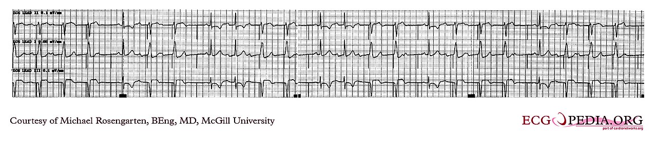
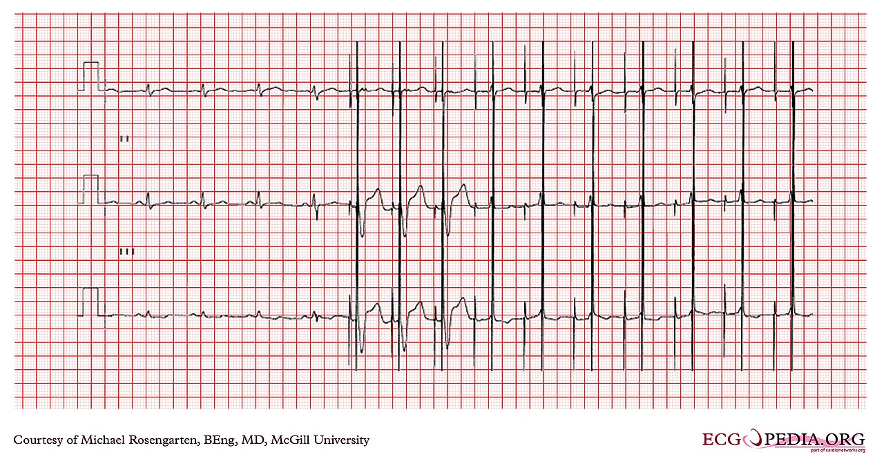
- Capture on some beats and not others: time-dependent failure (see Figure 1)
- A spike followed by an escape or native rhythm, not a paced complex
Causes
- Lead dislodgement, lead fracture or insulation failure, fibrosis or a rise in threshold
- Myocardial infarction or scar at the lead tip, which raises the threshold
- Electrolyte shifts that raise threshold, such as hyperkalemia or acidosis; hypoxia
- Drugs that raise threshold: class IC antiarrhythmics such as flecainide, and amiodarone
- Low battery or an output setting that is too low for the threshold
Clinical Significance
- Confirm capture by pulse and SpO₂ waveform. An unconfirmed spike is not pacing
- Without capture, the patient may be on an escape rhythm with a slow rate. That needs urgent device review
- See Ventricular Rhythms for the escape rhythm patterns
Failure to Sense: Undersensing and Oversensing
Sensing is how the device detects the patient's own beats. When sensing fails, the device either ignores a native beat or counts noise as a beat.
ECG Criteria
- Undersensing: pacing spikes fall on native QRS complexes or on T waves
- Oversensing: pacing pauses or drops out when the device counts noise or another signal as a beat
- Pacing spike timing that does not match the programmed rate or mode
Causes
- Lead dislodgement or a lead fault
- Electrical interference from the monitor, other devices or muscle artifact
- A sensing setting that is too sensitive or not sensitive enough for the patient
Clinical Significance
- Undersensing can put a spike on a T wave. Treat it as urgent and call the device team
- Oversensing can produce pauses. Check pulse and SpO₂ before you assume the rhythm changed
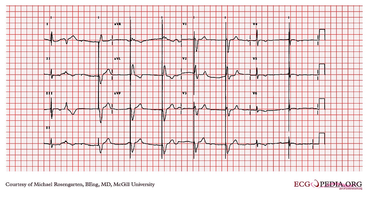
- Figure 2 shows a dual-chamber pacemaker strip used to confirm capture after a magnet was applied
Failure to Pace: Output Failure and Pauses
When the device goes quiet, or fires too late to stop a pause, that is failure to pace. Now the escape rhythm is all the patient has.
ECG Criteria
- No pacing spikes after a pause in a patient who is supposed to be paced
- Rate below the programmed lower rate with no spikes
- An escape rhythm with a wide QRS and slow rate, which needs urgent treatment
Causes
- Battery depletion or a device at elective replacement
- Lead fracture or dislodgement
- Output set too low for the current threshold
Clinical Significance
- Hypotensive, confused or with ischemic chest pain? Treat it as unstable bradycardia
- Transcutaneous pacing is the bridge: see Transcutaneous Pacing
- Send the device identification card and call the device clinic
Pacemaker-Mediated Tachycardia
Pacemaker-mediated tachycardia (PMT), also called endless-loop tachycardia, is a tracking loop. It starts with a premature ventricular beat, or with backward conduction from ventricle to atrium, that leaves behind a retrograde P wave. The device hears that P wave as a cue and paces the ventricle again. Around it goes, at the upper tracking rate.
ECG Criteria
- Regular tachycardia at the programmed upper tracking rate
- A pacing spike precedes each QRS; the retrograde P wave may be hidden in the T wave
- Onset after a premature beat, then self-sustaining
Management
- Treat it as a device problem and call the device team. In some pacemakers a magnet may break the loop, since it switches the device to fixed-rate pacing (device-specific)
- The definitive fix is reprogramming by the device team, such as a longer PVARP or a non-tracking mode
- In an ICD, a magnet usually does not stop the loop. It suspends therapies rather than changing the tracking
Magnet Response: Pacemaker vs ICD
Put a magnet on a pacemaker, then on an ICD, and you get two different results. Most pacemakers go asynchronous at a fixed rate, with sensing off. Most ICDs suspend tachy-therapies (ATP and shocks) and leave bradycardia pacing alone. The response varies with manufacturer, model and battery state. The device card or device record is the reference.
- Pacemaker: asynchronous pacing at a fixed rate. The magnet shows the device is pacing. It does not test capture
- ICD: tachy-therapies are suspended. Shock protection is gone while the magnet is in place
- Not a capture test: the magnet's pacing rate tells you nothing about whether each spike captures. Confirm capture by pulse and SpO₂ waveform, or have the device team check it with a programmer
Paced Rhythms: Ischemia & Morphology
A paced QRS is wide, and its ST segments run discordant. Standard STEMI criteria do not apply. Use the Smith-modified Sgarbossa criteria and compare with a prior 12-lead.
Smith-Modified Sgarbossa Criteria
- Concordant ST elevation of 1 mm or more in leads with a positive QRS
- Concordant ST depression of 1 mm or more in V1–V3
- Excessively discordant ST elevation: ST/S ratio of −0.25 or less, replacing the original 5 mm rule
Paced Morphology by Lead Site
- Right ventricular apical pacing: a wide, left bundle branch block-like QRS
- Biventricular (CRT) pacing: the QRS can be narrower than with RV pacing alone, and the V1 pattern can change
- Leadless and His or left bundle area pacing: morphology depends on the lead site and can be narrower
Judge a paced morphology against the prior 12-lead and the device record. See ST Segment & Ischemia.
Pacemaker Syndrome & Runaway Pacemaker
Pacemaker Syndrome
In a ventricular-paced patient without AV synchrony, the atria and ventricles contract at different times. Watch for symptoms such as dizziness, fatigue, hypotension and dyspnea. Look for cannon A waves or AV dissociation on the rhythm strip. Then refer to the device team for a mode change.
Runaway Pacemaker
A runaway pacemaker fires at a rapid rate. Electromagnetic interference or a lead fault may be the cause. A magnet sets fixed-rate pacing, and it may not slow the device. If there is an interference source, remove it, then call the device team urgently.
ICD Shocks & Electrical Storm
An ICD tries antitachycardia pacing (ATP) first. Shocks come if needed. Electrical storm can happen in ICD patients, and it carries high short-term mortality.
ECG Criteria
- Electrical storm: three or more episodes of sustained ventricular arrhythmia within 24 hours, at least 5 minutes apart, each needing ATP or a shock
- Inappropriate shocks: SVT, including atrial fibrillation with a rapid ventricular response, or lead noise and oversensing can trigger shocks that are not needed. Find them on the rhythm strip before you treat a shock as VT or VF. Treat the rhythm, not the device alarm
Causes
- Ventricular arrhythmia from scar, ischemia or electrolyte disturbance
- Sympathetic surge, pain or distress
- Inappropriate detection of supraventricular rhythms or noise
Clinical Significance
- Pulseless VT or VF: defibrillate; a shock is the treatment (see Defibrillation)
- VT with a pulse and instability: synchronized cardioversion per the tachycardia algorithm (see Cardioversion)
- Stable sustained VT with a pulse: amiodarone and a rate-appropriate plan through the tachycardia algorithm; do not rely on the ICD to end it
- ICD patients still receive external shocks when they are needed. The implanted device changes nothing about the external protocol
- ATP type: the device team programs burst and ramp ATP. Which one ends a given tachycardia depends on the device and the VT. Do not guess at settings at the bedside
- Electrical storm: it needs urgent specialist input. Go after the reversible triggers: correct potassium and magnesium, and treat ischemia and decompensated heart failure
- Drugs: a non-selective beta-blocker such as propranolol is preferred, unless hypotension, shock, severe bradycardia or decompensated heart failure rules it out. Amiodarone goes in with it. Deep sedation cuts sympathetic drive. Doses are on the Amiodarone page and the beta-blocker pages
- If no programmer is available, a magnet over an ICD can disable its therapies. That removes shock protection, so it is the device team's decision
Differential Diagnosis & Mimics
Start with table one to tell a real spike from artifact. Table two is the lineup of mimics, the things that pass for malfunction or for some other rhythm.
| Feature | Pacing spike | Artifact or interference |
|---|---|---|
| Timing | Fixed interval set by the device | Random or tied to movement |
| Shape | Brief, sharp, the same on each beat | Varies with motion; wanders across the strip |
| Follow-through | Capture produces a P wave or QRS | No following complex |
Device and ECG Mimics
| Mimic | What it looks like | Clue |
|---|---|---|
| Hysteresis | A pause after a native beat, slower than the programmed rate | Normal when programmed; check the device card and the device record |
| Rate response or sleep rate | A slow paced rate at rest or during sleep | Rate rises with activity; a lower sleep rate is programmed |
| Pseudofusion | A spike falls just before or within a native QRS | A fused complex whose shape changes with spike timing |
| Spike in the refractory period | A spike shortly after a native QRS with no paced complex | Normal inhibition; the chamber cannot respond. Device interrogation confirms it |
| VT vs paced wide complex | A wide QRS at a regular rate | Paced rhythm has a spike before each QRS; VT spikes do not precede every QRS. Compare with a prior 12-lead |
Artifact & Lead Misplacement
- Muscle tremor or shivering can look like spikes. Confirm by pulse
- Interference from the monitor or other equipment can create false spikes
- Poor electrode contact can hide a spike in one lead. Check a second lead
- Bipolar pacing spikes are small, and some monitors miss them. A paced rhythm can then read as asystole or bradycardia. Turn on pacer detection where the monitor offers it, and check a second lead
Bedside Management & Escalation
| Situation | BLS (EMT) | ALS (AEMT / Paramedic / RN) | Critical Care / Transport |
|---|---|---|---|
| Unstable bradycardia (no device failure) | Oxygen if SpO₂ is below 94%, notify ALS, 12-lead where allowed | Atropine 1 mg IV/IO (AHA bradycardia algorithm); if no response, transcutaneous pacing or a dopamine or epinephrine infusion | Transvenous pacing or infusion while the device team is called |
| Pacemaker failure, failure to capture, or wide-complex escape | Oxygen if SpO₂ is below 94%, notify ALS, do not delay transport | Go to transcutaneous pacing early; do not wait for atropine. Start a dopamine or epinephrine infusion as the bridge. Confirm capture by pulse or SpO₂ waveform | Transvenous pacing via the device team; keep pads on for TCP |
| Stable (paced, capture confirmed) | Monitor; do not interfere with the device | 12-lead; call device clinic; check capture | Device card with the patient; pads placed to allow pacing or shock |
| Pulseless (VF or pVT) | CPR and AED shock | Defibrillation without delay, then ALS algorithm. Do not hold CPR or the shock to interrogate a device | Defibrillate without delay; do not hold the shock for a device. Keep pads away from the generator; follow local protocol |
| Therapy | Adult | Pediatric | Key Notes |
|---|---|---|---|
| Atropine | 1 mg IV/IO every 3–5 min (max 3 mg) | 0.02 mg/kg IV/IO (min 0.1 mg, max 0.5 mg), may repeat once | Stop after 3 mg (adult). For bradycardia from increased vagal tone or primary AV block only; see Atropine. Unlikely to help infranodal block and not first-line for pacemaker failure |
| Dopamine | 5–20 mcg/kg/min IV infusion, titrated | See the E3 dopamine page | High-alert Pacing bridge; see Dopamine |
| Epinephrine | 2–10 mcg/min IV infusion | See the E3 epinephrine page | High-alert Alternative when pacing is delayed; see Epinephrine |
| Amiodarone | VF or pVT: 300 mg IV/IO, then 150 mg. Stable VT: 150 mg IV over 10 min | 5 mg/kg IV/IO (max 300 mg) | High-alert Do not give in torsades; see Amiodarone |
Resuscitation With a Device
- Defibrillate without delay. Do not hold the shock or CPR to interrogate or program a pacemaker or ICD.
- Pad placement: keep pads away from the generator. If the generator is in the way, use anterior-posterior placement, as the defibrillation page describes.
- Transcutaneous pacing: keep pads away from the generator. Confirm capture by pulse or SpO₂ waveform, not the monitor alone; pacer artifact can look like capture.
- After any shock: have the device interrogated by the device team.
- Pregnancy: standard ALS still applies, defibrillation included. Shocks and standard drugs are not withheld. Manual left uterine displacement comes into use after about 20 weeks' gestation.
Pacing: see Transcutaneous Pacing, Defibrillation and Cardioversion. Defibrillation: manufacturer biphasic dose (120–200 J) or 360 J monophasic.
Follow local protocols and online medical direction.
Pediatric & Special Populations
Pediatrics
- Children with congenital heart disease may have epicardial wires and an external generator. Settings come from cardiology or the surgical team
- Pediatric atropine and epinephrine doses are on the E3 pages. Weight-based dosing lives on Weight-Based Dosing
- Pediatric transcutaneous pacing: pad size and placement follow the pediatric protocol; confirm capture by pulse
- Pediatric ICD: cardiology or the device clinic sets the shock and ATP settings. External shock doses are weight-based (see Weight-Based Dosing)
- Vital sign ranges by age: Vital Signs
Other Populations
- Heart transplant: the heart is denervated, so atropine is unlikely to help bradycardia
- Pacemaker or ICD in place: identify the device type and read the card before any procedure. Magnet response is device-specific (see the Magnet Response section)
- Pregnancy: standard ALS still applies, defibrillation included (see Resuscitation With a Device). Review the device and its settings with cardiology
Monitoring & Transport Considerations
| Issue | What to Do |
|---|---|
| Best lead | Lead II for spikes and P waves; V1 for ventricular morphology |
| Capture | Confirm by pulse and SpO₂ waveform after each change in position or setting |
| Transport | Bring the device card; notify the receiving team that the patient is paced |
| Interference | Keep phones and other equipment away from the generator; watch for false spikes |
| Serial 12-leads | Repeat when the rhythm or chest pain changes; compare with a prior paced 12-lead |
Step-by-Step Approach & Pitfalls
- Confirm the patient: check pulse, SpO₂ and mental status before reading the strip.
- Find the spikes: look in lead II and V1 for every spike.
- Check capture: is each spike followed by a P wave or QRS?
- Check sensing: do any spikes fall on native beats or T waves?
- Check the rate: compare with the programmed lower rate on the device card.
- Act on the patient: treat unstable bradycardia per the table above and call the device team.
Common Pitfalls
- Calling a spike a QRS. A spike with no complex is not a beat
- Taking the monitor rate at face value, with no pulse check
- Applying standard STEMI criteria to a paced QRS. Use Sgarbossa and a prior 12-lead
- Giving atropine and waiting in Mobitz II or complete block with a wide escape
- Reprogramming the device at the bedside without the device team
ECG Examples
Clinical Pearls
References
- American Heart Association. Part 9: Adult Advanced Life Support: 2025 American Heart Association Guidelines for Cardiopulmonary Resuscitation and Emergency Cardiovascular Care. Circulation. 2025;152(16 suppl 2):S538–S577. https://doi.org/10.1161/CIR.0000000000001376
- 2018 ACC/AHA/HRS Guideline on the Evaluation and Management of Patients With Bradycardia and Cardiac Conduction Delay. Circulation. 2019;140. https://doi.org/10.1161/CIR.0000000000000628
- 2017 AHA/ACC/HRS Guideline for Management of Patients With Ventricular Arrhythmias and the Prevention of Sudden Cardiac Death. Circulation. 2018;138. https://doi.org/10.1161/CIR.0000000000000549
- 2022 ESC Guidelines for the management of patients with ventricular arrhythmias and the prevention of sudden cardiac death. Eur Heart J. 2022;43(40):3997–4126. https://doi.org/10.1093/eurheartj/ehac262
- Bernstein AD, Daubert JC, Fletcher RD, Hayes DL, Lüderitz B, Reynolds DW, Schoenfeld MH, Sutton R. The revised NASPE/BPEG generic code for antibradycardia, adaptive-rate, and multisite pacing. Pacing Clin Electrophysiol. 2002;25:260–264. https://doi.org/10.1046/j.1460-9592.2002.00260.x
- Chung MK, et al. 2023 HRS/APHRS/LAHRS guideline on cardiac physiologic pacing for the avoidance and mitigation of heart failure. Heart Rhythm. 2023;20(9):e17–e91. https://doi.org/10.1016/j.hrthm.2023.03.1538
- Smith SW, Dodd KW, Henry TD, Dvorak DM, Pearce LA. Diagnosis of ST-elevation myocardial infarction in the presence of left bundle branch block with the ST-elevation to S-wave ratio in a modified Sgarbossa rule. Ann Emerg Med. 2012;60(6):766–776. https://doi.org/10.1016/j.annemergmed.2012.07.119
Medical Disclaimer
- For Educational Purposes Only: This content is intended for educational reference and should not be used for clinical decision-making.
- Not a Substitute for Professional Judgment: Always consult your local protocols, institutional guidelines, and supervising physicians.
- Verify Before Acting: Users are responsible for verifying information through authoritative sources before any clinical application.