

At a Glance
- Represents
- Atrial depolarization, AV nodal delay and His–Purkinje conduction, measured from P-wave onset to QRS onset.
- Normal (Adult)
- 120–200 ms (0.12–0.20 s; 3–5 small boxes), the same on every conducted beat.
- Measure It
- Lead II: P-wave onset to earliest QRS onset. Check several beats for the shortest and longest PR.
- Abnormal → Think
- >200 ms: first-degree AV block; review drugs and potassium
- <120 ms: pre-excitation (delta wave, wide QRS) or junctional rhythm
- Variable PR: higher-degree AV block (see Conduction Blocks)
- Red Flags
- Short PR + delta wave with an irregular, wide, very rapid rhythm: pre-excited AF until proven otherwise.
- Bedside Action
- Pre-excited AF: no AV nodal blockers or IV amiodarone; unstable: synchronized cardioversion. Unstable bradycardia: atropine if nodal; go straight to pacing for wide-QRS or high-grade block.
Physiology & What It Represents
The PR interval starts at P-wave onset and ends at QRS onset. In that window the atria depolarize, the AV node holds the impulse back, and the signal travels the His–Purkinje system. Only then do the ventricles fire.
- Path: SA node, atria, AV node, His bundle, bundle branches, Purkinje fibers, ventricular myocardium
- Largest component: the AV node delay. It buys the atria time to finish before the ventricles contract
The AV node answers to autonomic tone and to AV nodal drugs, so the PR is your bedside read on AV conduction. A long PR points to slowed AV conduction. A short PR means the impulse reached the ventricles faster than the AV node should allow. Most often, an accessory pathway is the reason.
Normal Values & How to Measure
| Parameter | Adult Normal | Pediatric Note | Where / How to Measure |
|---|---|---|---|
| PR interval | 120–200 ms (0.12–0.20 s; 3–5 small boxes at 25 mm/s) | Age-dependent; use pediatric normal limits (see Pediatric section) | P-wave onset to earliest QRS onset, in a lead with clear P waves (usually II) |
| First-degree AV block | >200 ms (>5 small boxes) with 1:1 conduction | Evaluate with age-specific limits | Consistent PR on every conducted beat |
| Short PR | <120 ms (<3 small boxes) | Shorter PR is common in infants | Check for delta wave and QRS width |
| Rate dependence | PR shortens with faster rates and lengthens with slower rates | Interpret against heart rate | Compare PR with the rate on the same strip |
Measurement Steps
- Choose the lead: use a lead with a clear P wave and a clear QRS onset (usually lead II).
- Mark P-wave onset: the first deflection away from the baseline.
- Mark QRS onset: the earliest ventricular deflection, whether Q, R or S. In pre-excitation, measure to the start of the delta wave, not the peak of the QRS.
- Count: each small box is 40 ms (0.04 s). Count boxes or use calipers.
- Repeat on several beats: in a variable rhythm, record the shortest and longest PR. Confirm that every P wave conducts before you call it first-degree block.
Prolonged PR Interval (First-Degree AV Block)
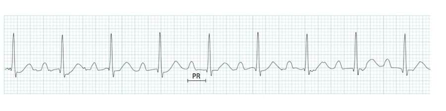
First-degree block means a PR longer than 200 ms (>5 small boxes). Conduction stays 1:1, so every P wave gets a QRS behind it.
ECG Criteria
- PR interval: >200 ms, constant from beat to beat
- Conduction: 1:1; no P wave is blocked
- QRS: usually narrow. The width helps you find a coexisting block
Causes
| Category | Examples |
|---|---|
| Medications | Beta-blockers, diltiazem, verapamil, digoxin, amiodarone, class IA and IC antiarrhythmics |
| Increased vagal tone | Athletes, sleep |
| Ischemia or infarction | Inferior MI (AV nodal supply), anterior MI (infranodal disease) |
| Inflammatory | Myocarditis, Lyme disease, rheumatic fever, endocarditis |
| Infiltrative or degenerative | Sarcoidosis, amyloidosis, age-related conduction fibrosis |
| Metabolic | Hyperkalemia, hypothyroidism |
| Congenital | Ebstein anomaly, atrial septal defect, neonatal lupus |
Clinical Significance
- Usually benign: most patients have no symptoms and need no AV-block-specific treatment. A very long PR (over 300 ms) can cause symptoms when the atria and ventricles lose sync. If it does, refer for evaluation.
- Drug review first: check AV nodal blockers, digoxin, amiodarone and electrolytes before you blame the block on disease
- Pacing: not indicated for a first-degree block without symptoms. If a very long PR causes symptoms, pacing may be considered after evaluation (2018 ACC/AHA/HRS). For higher-degree block, go to the Conduction Blocks page.
- Bifascicular block: add it to a first-degree block and the risk of complete heart block goes up. Trifascicular block is covered on the Conduction Blocks page.
Short PR Interval and Pre-Excitation (WPW)
A PR shorter than 120 ms means AV conduction is faster than the normal AV node delay. One of three things is behind it: a bypass tract (pre-excitation), enhanced AV nodal conduction, or a junctional rhythm without pre-excitation. Metabolic and storage diseases can shorten the PR too, including Fabry disease, Pompe disease and PRKAG2 syndrome.
| Condition | PR | QRS | Significance |
|---|---|---|---|
| Wolff-Parkinson-White (WPW) | <120 ms | Wide, with delta wave | Accessory pathway; risk of AVRT and AF with rapid conduction |
| Lown-Ganong-Levine (LGL) | <120 ms | Narrow, no delta wave | Enhanced AV conduction or a bypass tract; the existence of a true syndrome is debated |
| Junctional rhythm | <120 ms or absent | Narrow | Impulse arises near the AV node; retrograde P waves may be present |
Wolff-Parkinson-White (WPW) Syndrome
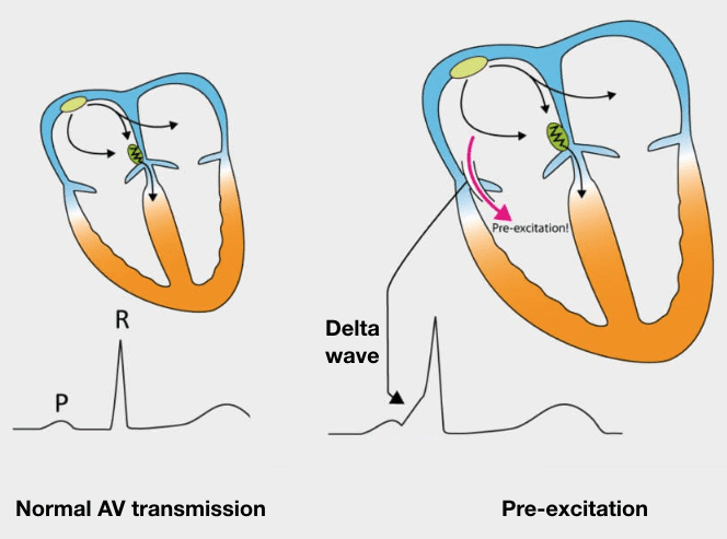
- Mechanism: an accessory pathway carries the impulse from atria to ventricles outside the AV node. That activates part of the ventricle early
- ECG features: PR <120 ms; wide QRS from fusion of normal and pre-excited conduction; slurred initial upstroke (delta wave); secondary ST-T changes opposite the QRS direction
- Arrhythmia risks: AVRT is the most common tachycardia in WPW. AF can conduct fast down the accessory pathway, and that can degenerate into ventricular fibrillation
Lown-Ganong-Levine (LGL) Pattern
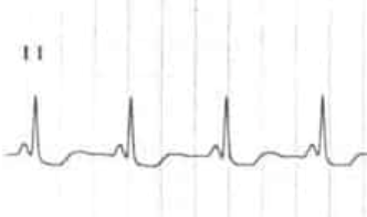
- Definition: PR <120 ms with a normal QRS and no delta wave
- Proposed mechanism: enhanced AV nodal conduction or a bypass tract (James fibers)
- Clinical view: many experts treat it as a variant. Whether it causes SVT is still unclear
Variable PR Interval
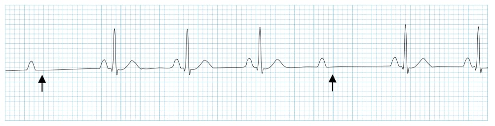
A PR that changes from beat to beat means AV conduction is intermittently variable. That pattern points to a higher-degree block. Mobitz I (Wenckebach) lengthens the PR a little more with each beat until a QRS drops. Mobitz II keeps the PR constant on the conducted beats, then drops a QRS without warning. The classification, the 2:1 problem and the pacing decisions are on the Conduction Blocks page.

Differential Diagnosis & Mimics
| Feature | WPW | LGL | Junctional rhythm |
|---|---|---|---|
| PR | <120 ms | <120 ms | <120 ms or absent |
| QRS | Wide (fusion) | Narrow | Narrow |
| Delta wave | Present | Absent | Absent |
| P waves | Sinus or AF; may be retrograde in AVRT | Sinus | Absent, or retrograde before/within/after QRS |
Artifact & Lead Misplacement
- Limb lead reversal: P waves in lead I flip upside down and the P-wave axis shifts. It can pass for a junctional or ectopic atrial rhythm
- Baseline wander or muscle tremor: hides the P-wave onset, so the PR can look longer or shorter than it is
- Hidden P waves: a P wave buried in the T wave or inside flutter waves changes the PR you think you see
- Transport vibration: the noise can pass for fibrillation or for changing P waves
Normal Variants
A PR of about 100–119 ms with a narrow QRS and no delta wave can be a normal variant. It is most common in young or athletic patients. Make sure a 12-lead shows no delta wave before you call it normal.
Bedside Management & Escalation
| Situation | BLS (EMT) | ALS (AEMT / Paramedic / RN) | Critical Care / Transport |
|---|---|---|---|
| Unstable bradycardia from AV block (hypotension, altered mental status, chest pain, dyspnea) | Oxygen if hypoxemic (protocol threshold; SpO₂ <94% is one common cutoff; no routine oxygen when normoxic); 12-lead if allowed; early ALS; apply pads | Atropine (table below) only if the block is nodal; go straight to transcutaneous pacing for wide-QRS or high-grade block, or if atropine fails; dopamine or epinephrine infusion while pacing is set up; notify cardiology | Pacing capture checks by pulse and SpO₂ waveform; transvenous pacing per receiving center |
| Stable first-degree AV block (PR prolonged, asymptomatic) | Monitor; 12-lead if allowed; notify ALS if symptoms appear | Review AV nodal and conduction-slowing drugs and potassium | Document PR; watch for progression to Mobitz II or complete block |
| Pre-excited AF (irregular, wide, very rapid QRS with short PR) | Pulseless: defibrillate per protocol. Stable: early ALS; do not give AV nodal blocking drugs or IV amiodarone | Unstable: synchronized cardioversion; cardioversion is preferred with any hemodynamic compromise. Stable: procainamide or ibutilide (per receiving center or EP) | Cardiology or electrophysiology consult; continuous monitoring |
Doses vary by protocol; follow local protocols and medical direction.
| Therapy | Adult | Pediatric | Key Notes |
|---|---|---|---|
| Atropine | 1 mg IV/IO every 3–5 min (max 3 mg) | 0.02 mg/kg IV/IO (min 0.1 mg, max 0.5 mg per dose; may repeat once) | Works at the nodal level only; often ineffective at infranodal block (Mobitz II, wide QRS). Do not delay pacing. In children, bradycardia is usually hypoxic: ventilate first; atropine is for increased vagal tone or primary AV block (see the pediatric epinephrine row). |
| Dopamine | 5–20 mcg/kg/min IV infusion, titrated (max 20 mcg/kg/min) | 2–20 mcg/kg/min IV infusion, titrated (per the E3 dopamine page) | High-alert Confirm mcg/kg/min vs mcg/min and the pump concentration. Extravasation is a risk: use a proximal IV or central access; phentolamine is used for infiltration (see the dopamine page). |
| Epinephrine bolus | Infusion only (see below) | 0.01 mg/kg (0.1 mL/kg of 0.1 mg/mL) IV/IO every 3–5 min (max 1 mg per dose) | High-alert First-line for pediatric bradycardia with poor perfusion, after oxygenation, ventilation and CPR if HR is below 60/min. Not the 1 mg adult arrest dose. |
| Epinephrine infusion | 2–10 mcg/min IV infusion, titrated | 0.1–1 mcg/kg/min IV/IO infusion, titrated (per the E3 epinephrine page) | High-alert Infusion is in mcg/min; do not confuse with the 1 mg arrest dose. |
| Transcutaneous pacing | Per transcutaneous pacing page | Per the same page (pediatric pad size and settings) | Anterior–posterior pads; confirm capture by pulse and SpO₂ waveform; sedation or analgesia as tolerated. |
| Procainamide (stable pre-excited AF) | Per the E3 procainamide page: 20–50 mg/min IV until the arrhythmia stops, BP falls, QRS widens more than 50%, or 17 mg/kg is reached | Expert consultation; see the E3 procainamide page | High-alert Contraindicated with 2nd- or 3rd-degree AV block without pacing, with torsades, and in established SLE. Avoid with prolonged QT or heart failure; monitor BP and QRS continuously. The 2023 AF guideline lists IV procainamide or ibutilide for stable pre-excited AF; dosing is protocol-dependent. |
| Synchronized cardioversion (unstable pre-excited AF) | Energy per cardioversion page | Energy per the same page | Sedate if time allows; do not delay for medication in an unstable patient. |
| AV nodal blockers and amiodarone | Avoid: adenosine (adenosine), diltiazem (diltiazem), verapamil (verapamil), beta-blockers (e.g., metoprolol), digoxin and IV amiodarone | Avoid | In pre-excited AF these drugs and IV amiodarone can accelerate the ventricular rate and precipitate VF. |
Follow local protocols and online medical direction.
Pediatric & Special Populations
Pediatrics
- Age-specific limits: the normal PR changes with age. For infants and children, use a pediatric reference such as the normal-limits study by Rijnbeek (2001). Adult limits do not apply to them.
- Pediatric bradycardia: ventilate and oxygenate first. If HR is below 60/min with poor perfusion despite that, start CPR. Epinephrine 0.01 mg/kg (0.1 mL/kg of 0.1 mg/mL) IV/IO is the first-line drug. Atropine (0.02 mg/kg) is for increased vagal tone or primary AV block. See the epinephrine and atropine pages.
- Heart rate: children have faster resting rates. Read the PR against their age-specific rate (see pediatric vital signs).
- First-degree AV block in children: before you blame conduction disease, look for congenital heart disease, myocarditis, digoxin toxicity and electrolyte abnormalities.
- Drug dosing: dose atropine, adenosine and antiarrhythmics by weight-based dosing.
Other Populations
- Athletes: high vagal tone can lengthen the PR. Judge it by symptoms and the rhythm pattern.
- Older adults: age-related conduction fibrosis makes a long PR more common.
- Heart transplant recipients: the sinus node has been denervated. That can make heart-rate responses to atropine and other drugs unreliable.
- Pacemaker or ICD: the AV delay in a paced rhythm is programmed, not intrinsic. Don't read it as conduction disease.
Monitoring & Transport Considerations
- Monitoring lead: lead II shows the P waves and the PR best. If you suspect a delta wave, confirm it on a 12-lead.
- Repeat 12-leads: get a fresh one when the PR changes, after an AV nodal drug dose, or when the rhythm changes.
- Artifact: vibration, shivering and movement in transport can hide P waves or look like AF. Check the rhythm at rest before you act on it.
- Pacing: place anterior–posterior pads before you move an unstable bradycardic patient. After each change, check capture by pulse and the SpO₂ waveform.
- Electrolytes: hyperkalemia can prolong the PR and widen the QRS. Send the potassium with the history when you have it.
- Handoff: tell the receiving team the PR range, any dropped beats, and any short-PR or pre-excitation pattern.
Step-by-Step Approach & Pitfalls
- Confirm conduction: check whether every P wave is followed by a QRS.
- Measure the PR: P-wave onset to earliest QRS onset, in lead II, on several beats.
- Classify the PR: normal (120–200 ms), prolonged (>200 ms), short (<120 ms) or variable.
- If prolonged with 1:1 conduction: first-degree AV block. Review drugs and electrolytes, and watch for progression.
- If short: look for a delta wave and a wide QRS. Ask about palpitations and check for AF.
- If variable or with dropped QRS: classify the block on the conduction-blocks page and assess stability before treating.
Common Pitfalls
- Measuring to the end of the P wave. That gives you the PR segment, not the PR interval.
- Calling a short PR benign before you look for a delta wave.
- Handing an AV nodal blocker to an irregular wide-complex tachycardia without thinking of pre-excited AF.
- Reflexively reaching for IV amiodarone on an irregular wide-complex tachycardia. Exclude pre-excited AF first. The 2023 AF guideline rates AV nodal blocking agents, including amiodarone, as Class 3: Harm in pre-excited AF.
- Calling a 2:1 block first-degree or Mobitz II from one PR value. You can't assess the PR on a 2:1 strip.
- Expecting atropine to fix an infranodal block.
Clinical Pearls
References
- Kusumoto FM, Schoenfeld MH, Barrett C, et al. 2018 ACC/AHA/HRS Guideline on the Evaluation and Management of Patients With Bradycardia and Cardiac Conduction Delay. Circulation. 2019;140:e382–e482. https://doi.org/10.1161/CIR.0000000000000628
- Joglar JA, Chung MK, Armbruster AL, et al. 2023 ACC/AHA/ACCP/HRS Guideline for the Diagnosis and Management of Atrial Fibrillation. Circulation. 2024;149:e1–e156. https://doi.org/10.1161/CIR.0000000000001193
- Lasa JJ, Dhillon GS, Duff JP, et al. Part 8: Pediatric Advanced Life Support: 2025 American Heart Association and American Academy of Pediatrics Guidelines for Cardiopulmonary Resuscitation and Emergency Cardiovascular Care. Circulation. 2025;152(16 suppl 2):S479–S537. https://doi.org/10.1161/CIR.0000000000001368
- Wigginton JG, Agarwal S, Bartos JA, et al. Part 9: Adult Advanced Life Support: 2025 American Heart Association Guidelines for Cardiopulmonary Resuscitation and Emergency Cardiovascular Care. Circulation. 2025;152(16 suppl 2):S538–S577. https://doi.org/10.1161/CIR.0000000000001376
- Rijnbeek PR, Witsenburg M, Schrama E, Hess J, Kors JA. New normal limits for the paediatric electrocardiogram. Eur Heart J. 2001;22(8):702–711. https://doi.org/10.1053/euhj.2000.2399
Medical Disclaimer
- For Educational Purposes Only: This content is intended for educational reference and should not be used for clinical decision-making.
- Not a Substitute for Professional Judgment: Always consult your local protocols, institutional guidelines, and supervising physicians.
- Verify Before Acting: Users are responsible for verifying information through authoritative sources before any clinical application.