

At a Glance
- Represents
- Ventricular repolarization: the recovery phase after the QRS, which reflects perfusion, ion balance and autonomic tone.
- Normal (Adult)
- Upright with an upright QRS, asymmetric with a gentle upslope; inverted in aVR; V1 may be flat, upright or inverted.
- Measure It
- Compare T height and shape with the QRS in the same lead (II or V2–V4), then check contiguous leads for the territory.
- Abnormal → Think
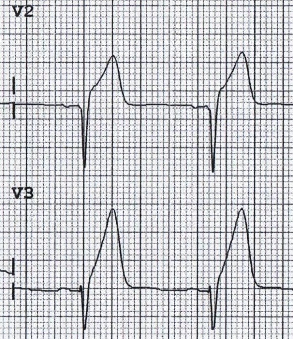
- Broad, tall, one territory: hyperacute T (occlusion MI)
- Narrow, peaked, diffuse: hyperkalemia
- Inverted or biphasic V2–V3: Wellens pattern
- Flat with prominent U waves: hypokalemia
- Red Flags
- Hyperacute T with chest pain or a matching territory: occlusion MI until proven otherwise. Peaked T with wide QRS, PR prolongation or lost P waves: hyperkalemia emergency.
- Bedside Action
- Hyperacute T: treat as possible occlusion MI; STEMI or OMI pathway per protocol now. Peaked T with ECG changes: IV calcium now, then insulin with dextrose. Wellens: urgent cardiology.
Physiology & What It Represents
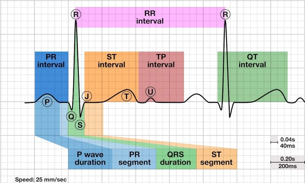
The T wave is ventricular repolarization. Each myocyte resets its resting membrane potential after it fires. That reset runs slower and less synchronized than the depolarization that made the QRS. So the T wave ends up smaller, broader and more variable than the QRS. Its direction normally follows the dominant QRS deflection.
Several things shape repolarization, and the T wave reflects every one of them:
- Regional action-potential duration: epicardial cells repolarize before endocardial cells. That timing sets the normal T-wave direction.
- Autonomic tone: sympathetic activity shortens repolarization. Vagal tone lengthens it.
- Ion gradients: shifts in potassium, calcium and magnesium change repolarization directly.
- Perfusion and oxygenation: ischemia changes the action potential early, before depolarization changes show up.
- Structure: hypertrophy and fibrosis change the order in which the heart repolarizes.
Normal Values & How to Measure
| Parameter | Adult Normal | Pediatric Note | Where / How to Measure |
|---|---|---|---|
| Direction | Concordant with the dominant QRS in most leads | Inverted T in V1–V3 is a common juvenile variant | Lead by lead, against the QRS in the same lead |
| Lead aVR | Inverted (its QRS is also negative) | Same | Expected; not an abnormality |
| Lead V1 | Upright, flat or inverted | Inversion more common in children | Interpret with V2; V1 is the most variable lead |
| Shape | Asymmetric: gentle upslope, steeper downslope | Same | Look at the best-visualized lead in each territory |
| Amplitude | Judged against the QRS in the same lead; no fixed cut-off on this page | Judged against the QRS and age | Compare with the R wave and with a prior ECG |
Measure the T wave with these steps:
- Find the T wave after the ST segment and note where it returns to baseline.
- Compare its direction with the QRS in the same lead.
- Judge its height against the QRS in that lead, not by absolute size alone.
- Note the shape: asymmetric and normal, broad-based, or narrow and tent-shaped.
- Check contiguous leads to find the distribution: anterior V1–V4, lateral I, aVL and V5–V6, or inferior II, III and aVF.
- Compare with a prior ECG. New T-wave change matters more than unchanged chronic change.
- Repeat the ECG after a few minutes if the rhythm, potassium or symptoms change. Serial tracings show evolution.
Hyperacute T Waves (Early Occlusion MI)
A hyperacute T wave is a tall, broad T wave in the territory of an acute coronary occlusion. It is one of the earliest ECG signs of occlusion MI (OMI), and it can show up before ST elevation does. Think of it as an early sign, not a diagnosis by itself. Look for ST changes and reciprocal depression alongside it.
ECG Criteria
- Shape: broad-based, tall and often symmetric; the QRS may lose amplitude in the same leads
- Distribution: confined to one territory, matching the suspected occluded artery
- Reciprocal change: ST depression in the opposite territory supports ischemia
- Timing: may precede ST elevation, and ST elevation may be small or absent in early OMI
Causes
- Acute coronary occlusion: the cause that matters. Map the territory onto the coronary anatomy it implies.
- Look-alikes: hyperkalemia and other tall T waves. Sort them out under Differential Diagnosis & Mimics.
Clinical Significance
- Treat a hyperacute T wave with ongoing symptoms as a possible occlusion MI until proven otherwise. It is not a formal STEMI criterion. Manage it as a STEMI equivalent per local protocol, and don't wait for ST elevation to appear.
- STEMI Equivalents & Occlusion MI Patterns covers the STEMI equivalents, including the de Winter pattern. The Wellens pattern lives on ST Segment & Ischemia.
Peaked T Waves and Hyperkalemia
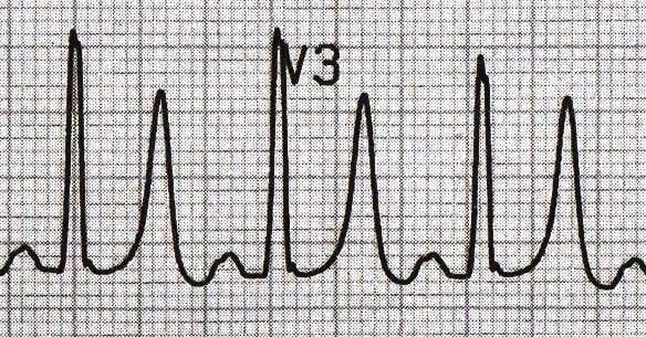
Peaked T waves are often the first ECG change in hyperkalemia. Read them as a warning, not a potassium level. The T wave is a poor ruler for the number.
ECG Criteria
- Shape: tall, narrow-based, symmetric and tent-shaped
- Distribution: diffuse, across leads that do not share one coronary territory
- Progression: peaked T waves, then P-wave flattening and PR prolongation, then QRS widening, a sine-wave pattern, and finally ventricular fibrillation, asystole or PEA
- Reliability: in a retrospective hospital series, the ECG was insensitive for hyperkalemia, and no T-wave threshold predicted it reliably. A normal ECG does not exclude hyperkalemia.
Causes
- Kidney failure: the kidneys shed less potassium. It is the most common setting.
- Medications: several drugs raise potassium, including ACE inhibitors and ARBs, potassium-sparing diuretics and some others, alone or in combination.
- Shifts and load: acidosis, cell breakdown and potassium-rich fluids
Clinical Significance
- Peaked T with QRS widening, PR prolongation or lost P waves is an emergency. Give IV calcium now. It protects the myocardium, but it does not lower potassium.
- Confirm with a serum potassium on a non-hemolyzed sample. Start potassium-shifting therapy while the result is pending.
- Other electrolyte patterns, including hypokalemia and the calcium and magnesium effects, are on Electrolyte Abnormalities on the ECG.
T-Wave Inversion
T-wave inversion is common. It runs from a normal variant all the way to acute ischemia. The territory, the symmetry and the comparison with a prior ECG decide what you do.
ECG Criteria
- Ischemic: symmetric, often deep, in a coronary territory, and new compared with a prior ECG
- Strain: asymmetric with a slow downslope and rapid upslope, often with ST depression: lateral leads in LV strain, V1–V4 in RV strain
- Cerebral: deep, widely distributed inversion with QT prolongation during intracranial hemorrhage or stroke
Causes
| Category | Examples |
|---|---|
| Ischemia | Acute or evolving MI; critical proximal LAD stenosis (see Wellens Pattern) |
| Strain | LV hypertrophy; RV pressure overload from acute pulmonary embolism |
| Cardiomyopathy | Hypertrophic cardiomyopathy (deep lateral or inferior inversion); Takotsubo |
| Central nervous system | Subarachnoid or intracerebral hemorrhage, ischemic stroke |
| Other | Bundle branch block (discordant T waves); late pericarditis; juvenile pattern in V1–V3; digoxin effect |
Clinical Significance
- Chest pain plus new T-wave inversion in contiguous leads means acute coronary syndrome until you rule it out.
- Diffuse inversion across most leads, with aVR left out: consider pulmonary embolism, myocarditis, Takotsubo or a CNS event.
- Cerebral T waves can be a CNS finding, not coronary ischemia. Myocardial injury can come along with them.
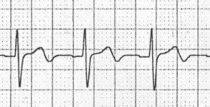
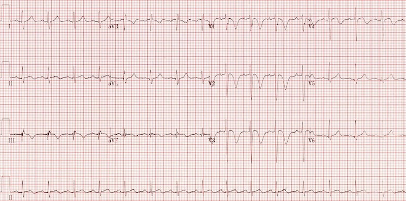
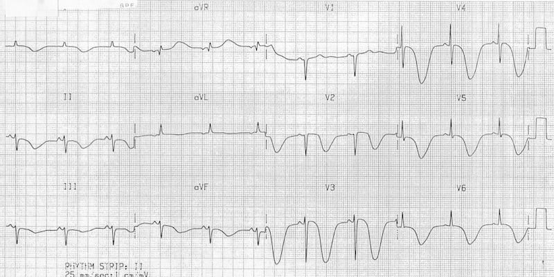
Wellens Pattern (Critical LAD Stenosis)
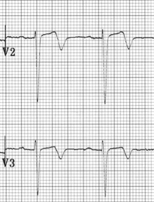
A Wellens pattern is a T-wave pattern in V2–V3 that signals critical proximal LAD stenosis. Full teaching lives on ST Segment & Ischemia. This page covers the T-wave view.
ECG Criteria
- Type A: biphasic T waves in V2–V3
- Type B: deeply inverted, symmetric T waves in V2–V3, often extending to V4
- Context: little or no ST elevation. Typically the chest pain has settled by then, though the pattern can already be present during an attack.
Clinical Significance
- The original 1989 series looked at angiography in patients with this pattern. Every one of them had at least 50% proximal LAD narrowing, and some had a total occlusion. The ECG changes were already there on admission in 108 of the 180 patients.
- Treat it as a pre-infarction pattern. That means urgent cardiology review and angiography per protocol. Never file it as a normal or benign T wave.

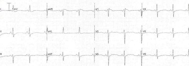
Flat T Waves and U Waves
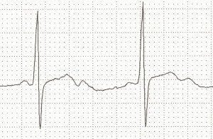
T-wave flattening tells you little on its own. In the electrolyte setting it points to hypokalemia, especially when U waves stand out and the QT interval looks prolonged.
ECG Criteria
- Flat T waves: low amplitude, close to baseline, in several leads
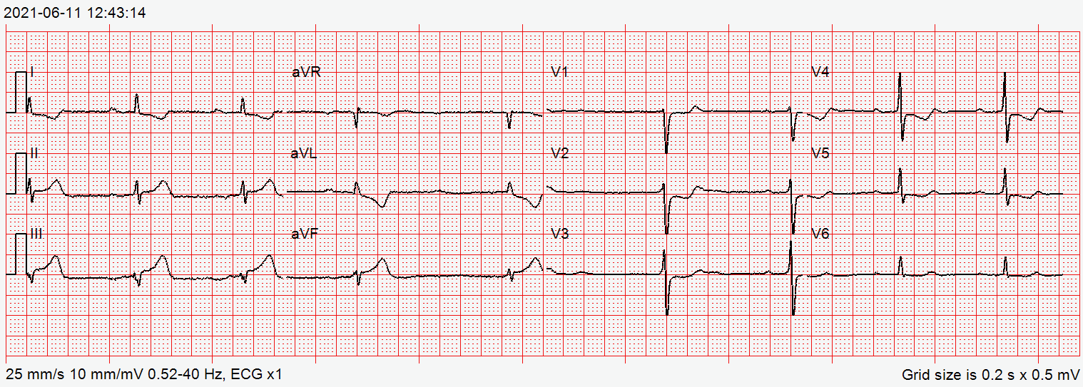
- U waves: small deflections after the T wave, most visible in V2–V3; prominent U waves suggest hypokalemia
- Apparent QT prolongation: a prominent U wave can fuse with the T wave and lengthen the apparent QT; measure carefully (see QT Interval)
Causes
- Hypokalemia: often the earliest ECG sign. As it worsens, the T wave progresses to inversion with prominent U waves.
- Hypomagnesemia: on the ECG it looks like hypokalemia, and the two often come together.
- Ischemia: subendocardial ischemia can flatten T waves or invert them only slightly.
- Low voltage: hypothyroidism, pericardial effusion and cardiomyopathy
- Normal variant: you see it often in older adults and in people with a thick chest wall.
T-Wave Alternans
T-wave alternans means the T wave changes from beat to beat in amplitude, polarity or shape. It reflects unstable repolarization, and it is linked to ventricular arrhythmia risk.
ECG Criteria
- Microvolt alternans: needs specialized equipment; it has been studied as a predictor of ventricular arrhythmia
- Macroscopic alternans: visible on a standard ECG, a sign of marked electrical instability
Causes
- Acute ischemia
- Long QT syndrome
- Severe electrolyte disturbance
Clinical Significance
- A patient with macroscopic alternans and any of ischemia, ventricular ectopy or a long QT needs immediate ED or ICU evaluation. Keep them on continuous monitoring for ventricular tachycardia and fibrillation.
Differential Diagnosis & Mimics
Tall or territorial T waves have several mimics. Going feature by feature, the hyperacute T and the hyperkalemic T do not look alike.
| Feature | Hyperacute T (OMI) | Hyperkalemia |
|---|---|---|
| Shape and base | Broad-based, tall, often symmetric | Narrow-based, tent-shaped (peaked) |
| Distribution | One coronary territory | Diffuse, across leads |
| Other ECG findings | Reciprocal ST depression; ST elevation may be small or absent early | PR prolongation, P-wave flattening, QRS widening as potassium rises |
| Clinical context | Chest pain, risk factors, ongoing symptoms | Kidney failure, potassium-raising drugs, acidosis, cell breakdown |
Look-Alikes to Recognize
- Left ventricular hypertrophy: tall lateral T waves that come with LVH voltage. See R Wave.
- Early repolarization: J-point elevation with notching. The STEMI mimics are on ST Segment & Ischemia.
- Athletes: symmetric, gradual T waves with no ischemic symptoms.
- Early pericarditis: tall T waves that appear before widespread ST elevation.
- Brugada pattern: T-wave inversion in V1–V2 with coved ST elevation
- Hypothermia: J (Osborn) waves with T-wave change
Artifact & Lead Misplacement
- Limb-lead reversal: flips T-wave direction in the affected leads. Left-right arm reversal, for example, inverts lead I.
- Precordial placement: electrodes placed high or low alter the T waves in V1–V3.
- Motion and tremor: shivering or movement can mimic peaked or wandering T waves. Repeat the tracing before you act.
Normal Variants
- Memory T waves: a brief T-wave inversion after tachycardia, ventricular pacing or bundle branch block. It is benign, yet it can mimic ischemia.
- Juvenile pattern: inverted T waves in V1–V3 in children and young adults
Bedside Management & Escalation
UKKA 2026 sets the adult non-arrest hyperkalemia doses, and AHA 2025 Part 10 sets arrest dosing. The linked E3 medication pages carry the dosing details.
| Situation | BLS (EMT) | ALS (AEMT / Paramedic / RN) | Critical Care / Transport |
|---|---|---|---|
| Hyperacute T, suspected OMI | 12-lead acquisition and transmission where allowed; oxygen if SpO₂ <94%; early notification | Repeat the 12-lead; aspirin and anticoagulation per ACS protocol after checking contraindications; notify the PCI center | Activate the cath lab; serial 12-leads every 15–30 min; right-sided and posterior leads |
| Peaked T, suspected hyperkalemia | 12-lead and monitor; ask about kidney failure and potassium-raising drugs; transport | IV calcium first if ECG changes are present; then insulin with dextrose; sodium bicarbonate only for severe acidosis (pH under 7.2, Paramedic/RN); repeat the ECG | Serial potassium and ECG; treat arrhythmia; monitor glucose for hours after insulin |
| New T inversion with chest pain (Wellens) | 12-lead and notification; oxygen if SpO₂ <94% | ACS protocol; aspirin per protocol; notify the cardiology center | Urgent cardiology and angiography; watch for evolution to STEMI |
| Pulseless, hyperkalemia suspected | CPR and defibrillation for VF per BLS | ALS arrest algorithm. AHA 2025 Part 10 (hyperkalemic arrest): IV calcium COR 2b, LOE C-LD; sodium bicarbonate COR 2b, C-EO; insulin with glucose COR 2b, C-EO (effectiveness not well established in each case); inhaled beta-2 agonists COR 3: No Benefit, C-EO. Suspected hyperkalemic arrest: calcium chloride 1 g IV/IO push; insulin 10 units with D50 50 g; albuterol not used. | Treat the cause; dialysis or potassium removal as the setting allows |
| Therapy | Adult | Pediatric | Key Notes |
|---|---|---|---|
| Calcium chloride 10% (Calcium Chloride) | 1 g (10 mL of 10%) IV/IO push in arrest; 10 mL over 5 min peri-arrest or with central/deep access; calcium gluconate 3 g (30 mL) over 10 min is the default for non-arrest patients; repeat if ECG changes persist No fixed max: check ionized calcium after repeated doses. Differs from FDA label: the US labels list only hypocalcemia and cap the rate (calcium gluconate 200 mg/min for adults, so 3 g takes at least 15 min; calcium chloride 1 mL/min, central or deep vein, no bolus). The hyperkalemia rate here (gluconate 30 mL over 10 min, chloride 10 mL over 5 min) is from UKKA 2026 and is off-label. | 20 mg/kg (0.2 mL/kg of 10%) IV/IO slowly (push in arrest), or gluconate 60 mg/kg (0.6 mL/kg of 10%) Max 1 g per dose (chloride); max 3 g per dose (gluconate) | Stabilizes the myocardium; does not lower K⁺. Label: give into a central or deep vein; extravasation causes tissue necrosis and calcinosis, so confirm patency. Label: avoid with digoxin; if it cannot be avoided, monitor ECG and calcium closely. Start K⁺-shifting therapy at the same time. In arrest from suspected hyperkalemia, AHA 2025 Part 10 rec 1 gives COR 2b, LOE C-LD (effectiveness not well established). Pediatric calcium doses (chloride 20 mg/kg, max 1 g; gluconate 60 mg/kg, max 3 g) are not stated in AHA 2025 or the FDA label and are not verified in a primary source. |
| Regular insulin with dextrose (Insulin; Dextrose) | 10 units IV with dextrose 25 g (non-arrest) Single dose; a second dose only if K⁺ is not below 6.0 within 2 hr, since each repeat adds hypoglycemia risk | 0.1 units/kg IV over 15–30 min with dextrose 0.5 g/kg (D10 5 mL/kg) Max 10 units per dose | High-alert Shifts K⁺ into cells. Always give the dextrose; if pre-dose glucose is under 126 mg/dL, add D10 50 mL/hr for 5 hr. Check glucose before the dose, then every 30 min for 2 hr, then hourly, for at least 6 hr. Hypoglycemia risk is higher in kidney failure, low body weight and low pre-dose glucose. Some US protocols use 5 units; UKKA 2026 says more study is needed before 5 units can be recommended, so this page uses 10 units for every adult dose. In arrest from suspected hyperkalemia, AHA 2025 Part 10 Table 3 gives 10 units with dextrose 50 g IV bolus (D50W) over 15–30 min; children get 0.1 units/kg (max 10 units) with D10W 5 mL/kg. Recommendation: COR 2b, LOE C-EO (effectiveness not well established). Differs from UKKA 2026 and ERC: those give 25 g (plus D10 50 mL/hr if glucose is under 126 mg/dL). |
| Sodium bicarbonate (Sodium Bicarbonate) | Not routine outside arrest (see Key Notes). In arrest: 50 mEq (one 50 mL vial) IV/IO (Paramedic/RN) Label (cardiac arrest): 44.6–100 mEq initially, then 44.6–50 mEq every 5–10 min as guided by blood gas. Repeat as guided by blood gas and pH | 1 mEq/kg IV/IO, slowly Max 50 mEq per dose. Label: 4.2% may be preferred in neonates and children under 2 years, because rapid hypertonic injection risks hypernatremia and intracranial hemorrhage | Shifts K⁺ into cells. Not routine outside arrest: consider only with severe metabolic acidosis (pH under 7.2) or when calcium is contraindicated. In suspected hyperkalemic arrest: 50 mEq IV/IO, repeat every 5–10 min guided by blood gas. AHA 2025 Part 10 rec 2 gives COR 2b, LOE C-EO (effectiveness not well established). It removes no potassium and has little potassium-lowering effect without acidemia. Label: do not add to calcium-containing solutions (precipitation); flush the line between drugs. |
| Albuterol, nebulized (Albuterol) | 10–20 mg nebulized (10 mg only with ischemic heart disease); add-on to calcium and insulin with dextrose, never alone Differs from FDA label: label nebulized dose is 2.5 mg for asthma; 10–20 mg is off-label, per UKKA 2026. | Per pediatric protocol; studies in ages 5–18 used 2.5–5 mg nebulized | Shifts K⁺ as an adjunct only; not a substitute for calcium or insulin. In arrest from suspected hyperkalemia, AHA 2025 Part 10 rec 4 gives inhaled beta-2 agonists COR 3: No Benefit, LOE C-EO; they are not recommended. |
| Potassium replacement (hypokalemia) | Oral or IV replacement per protocol with monitoring | Per pediatric protocol | High-alert Concentrated IV potassium: must be diluted; pump only, never IV push or undiluted. Label (Pfizer potassium chloride for injection concentrate, USP; see References): if serum K is above 2.5 mEq/L, give no faster than 10 mEq/h, in a final concentration of up to 40 mEq/L, with a 24-hour total not above 200 mEq. The 40 mEq/h urgent rate applies only when serum K is under 2 mEq/L with ECG changes and/or muscle paralysis, with continuous cardiac monitoring; it does not apply to other hypokalemia. |
Follow local protocols and online medical direction.
Pediatric & Special Populations
Pediatrics
- Juvenile T-wave pattern: inverted T waves in V1–V3 are a normal variant through childhood and adolescence. Before you call them ischemic, compare with prior ECGs and the clinical picture.
- Ventricular pacing: T waves that run against a paced QRS are expected. On their own they do not indicate ischemia.
- Pediatric dosing: calcium, insulin and bicarbonate scale with weight. Go to Weight-Based Dosing and check the per-dose maximum.
Other Populations
- Kidney failure and dialysis: New T-wave change calls for an urgent potassium check. Serial ECGs between dialysis sessions matter too.
- Older adults: low-amplitude or flat T waves can be nonspecific. Compare them with prior tracings.
- Pregnancy and postpartum: new T-wave change still gets the ACS evaluation. Keep peripartum cardiomyopathy on the differential.
- Athletes: tall, symmetric T waves without symptoms are often physiologic. Judge the morphology alongside the clinical picture.
Monitoring & Transport Considerations
- Monitor lead: pick the lead with the clearest T wave so you can trend changes. Use V2–V4 for anterior ischemia and II, III or aVF for inferior ischemia.
- Serial 12-leads: repeat every 15–30 minutes while chest pain persists or potassium-shifting therapy is running.
- Extra leads: add right-sided V4R when RV involvement is suspected, and posterior V7–V9 when posterior extension is in play. Do it where the protocol allows.
- Transmission: send the 12-lead early, where the protocol allows it. Note the time on every tracing.
- Artifact: shivering, vibration and movement in the ambulance can mimic peaked or flattened T waves. Check electrode contact and repeat the tracing before you act.
- Trends: the ECG tracks change. It is not the only basis for potassium treatment, so follow serial potassium values.
Step-by-Step Approach & Pitfalls
- Check direction: confirm the T wave is concordant with the QRS in the leads where the QRS is upright, and note aVR.
- Compare height with the QRS: judge the T wave against the QRS in the same lead, not in isolation.
- Describe the shape: broad-based and tall, narrow and tent-shaped, deeply inverted and symmetric, or biphasic.
- Map the distribution: one coronary territory, or diffuse across leads.
- Look for associated findings: ST elevation or depression, reciprocal change, PR and QRS changes, lost P waves, U waves and the QT interval.
- Compare with a prior ECG and repeat the tracing if the picture is unclear or changing.
- Act on the pattern: OMI pathway for a territorial hyperacute T; calcium and a potassium check for a peaked T; urgent cardiology for a Wellens pattern or new inversion with symptoms.
Common Pitfalls
- Waving off a hyperacute T wave because there is no ST elevation.
- Blaming hyperkalemia for every tall T wave, without checking the territory or the QRS.
- Waiting on a potassium result when peaked T waves come with QRS or PR changes.
- Reading a single ECG in an evolving patient. Serial tracings show the pattern.
- Mistaking a juvenile T-wave pattern in V1–V3 for a Wellens pattern, without checking the history.
- Missing limb-lead reversal. It inverts or changes T waves in the affected leads.
Clinical Pearls
References
- Rao SV, et al. 2025 ACC/AHA/ACEP/NAEMSP/SCAI guideline for the management of patients with acute coronary syndromes. Circulation. 2025;151:e771–e862. https://doi.org/10.1161/CIR.0000000000001309
- Cao D, et al. Part 10: Adult and pediatric special circumstances of resuscitation: 2025 American Heart Association guidelines for cardiopulmonary resuscitation and emergency cardiovascular care. Circulation. 2025;152:S578–S672. https://doi.org/10.1161/CIR.0000000000001380. AHA guidelines page: https://cpr.heart.org/en/resuscitation-science/cpr-and-ecc-guidelines/adult-and-pediatric-special-circumstances-of-resuscitation
- Rautaharju PM, et al. AHA/ACCF/HRS recommendations for the standardization and interpretation of the electrocardiogram: part IV: the ST segment, T and U waves, and the QT interval. J Am Coll Cardiol. 2009;53:982–991. https://doi.org/10.1016/j.jacc.2008.12.014
- Wagner GS, et al. AHA/ACCF/HRS recommendations for the standardization and interpretation of the electrocardiogram: part VI: acute ischemia/infarction. J Am Coll Cardiol. 2009;53:1003–1011. https://doi.org/10.1016/j.jacc.2008.12.016
- Montague BT, Ouellette JR, Buller GK. Retrospective review of the frequency of ECG changes in hyperkalemia. Clin J Am Soc Nephrol. 2008;3:324–330. https://doi.org/10.2215/CJN.04611007
- de Zwaan C, Bär FW, Janssen JH, et al. Angiographic and clinical characteristics of patients with unstable angina showing an ECG pattern indicating critical narrowing of the proximal LAD coronary artery. Am Heart J. 1989;117:657–665. https://doi.org/10.1016/0002-8703(89)90742-4
- de Winter RJ, Verouden NJ, Wellens HJ, Wilde AA. A new ECG sign of proximal LAD occlusion. N Engl J Med. 2008;359:2071–2073. https://doi.org/10.1056/NEJMc0804737
- Sovari AA, Assadi R, Lakshminarayanan B, Kocheril AG. Hyperacute T wave, the early sign of myocardial infarction. Am J Emerg Med. 2007;25:859.e1–859.e7. https://doi.org/10.1016/j.ajem.2007.02.005
- Rosenbaum DS, et al. Electrical alternans and vulnerability to ventricular arrhythmias. N Engl J Med. 1994;330:235–241. https://doi.org/10.1056/NEJM199401273300402
- UK Kidney Association. (2026). Clinical practice guideline: Management of hyperkalaemia in adults (updated July 2026). https://www.ukkidney.org/sites/default/files/documents/FINAL%20VERSION%20-%20UKKA%20CLINICAL%20PRACTICE%20GUIDELINE%20-%20MANAGEMENT%20OF%20HYPERKALAEMIA%20IN%20ADULTS%20-%20UPDATED%20JULY%202026_0.pdf
- Pfizer. Potassium Chloride for Injection Concentrate, USP: dosage and administration (prescribing information). Accessed 2026-10-15. https://www.pfizermedical.com/potassium-chloride/dosage-admin
Medical Disclaimer
- For Educational Purposes Only: This content is intended for educational reference and should not be used for clinical decision-making.
- Not a Substitute for Professional Judgment: Always consult your local protocols, institutional guidelines, and supervising physicians.
- Verify Before Acting: Users are responsible for verifying information through authoritative sources before any clinical application.