

Bedside Snapshot
- Normal (Adult)
- 1.12–1.32 mmol/L (4.5–5.3 mg/dL)
Whole blood on a blood-gas or point-of-care analyzer; typical, lab-specific - Critical Values
- <0.8 or >1.6 mmol/L (<3.2 or >6.4 mg/dL)
Typical; lab-specific - High Suggests
- Acidosis (lower pH raises iCa)
- Hypercalcemia from any cause
- Excess IV calcium or calcium supplements
- Low Suggests
- Citrate in massive transfusion (fast rates, liver dysfunction)
- Alkalosis, including hyperventilation or bicarbonate
- Sepsis, pancreatitis, rhabdomyolysis, hypomagnesemia
- Spurious: liquid heparin, hemolysis
- Act Now If
- iCa <0.9 mmol/L during transfusion, or low iCa with tetany, seizure or arrhythmia: give calcium chloride 1 g IV (or gluconate 3 g), monitor, recheck iCa
- Watch Out For
- Routine coagulation tests do not show the effect of low iCa. Measure iCa directly and recheck after each dose.
Reference & Critical Ranges
Reference ranges
| Population | Conventional | SI | Notes |
|---|---|---|---|
| Adult, ionized calcium | 4.5–5.3 mg/dL | 1.12–1.32 mmol/L | Whole blood; the active fraction. The 2023 European trauma guideline states the normal range as 1.1–1.3 mmol/L |
| Older adult | Same as adult | Same as adult | No age-specific limit verified for this page |
| Pregnancy | — | — | No pregnancy-specific range verified; use the laboratory range and the obstetric hemorrhage protocol |
| Neonates and children | Laboratory age-specific range | — | Not verified for this page; use the pediatric reference on your report |
Severity & critical values
| Level | Conventional | SI | What it means |
|---|---|---|---|
| Mild hypocalcemia | Below 4.4 mg/dL | <1.1 mmol/L | Trauma meta-analysis cut-off; higher mortality in observational data. Check the trend and the cause |
| Transfusion-induced hypocalcemia | Below 3.6 mg/dL | <0.9 mmol/L | Correct promptly during transfusion (2023 European guideline). If iCa is unavailable, a total corrected calcium of 7.5 mg/dL or lower is the guideline fallback; corrected calcium does not replace a measured iCa |
| Dysrhythmia risk | Below 3.2 mg/dL | <0.8 mmol/L | Linked to cardiac dysrhythmias in the 2023 European guideline: treat now and monitor |
| High | Above 5.3 mg/dL | >1.32 mmol/L | Above range: check total calcium and PTH; acidosis raises iCa, so read it with the pH. After calcium dosing, a high value means stop and recheck |
| Critical | Below 3.2 or above 6.4 mg/dL | <0.8 or >1.6 mmol/L | Typical panic values (lab-specific): the lab calls. Low: assess, monitor and treat now. High: stop further calcium, repeat the sample and treat the cause |
Conversion: iCa mg/dL × 0.25 = mmol/L (mmol/L × 4 = mg/dL); mmol/L × 2 = mEq/L
Causes of Abnormal Results
Low (below 1.12 mmol/L)
In trauma and massive transfusion, low iCa is mostly citrate chelation: the citrate in transfused products binds calcium. The effect is bigger when transfusion runs fast, when the liver cannot clear citrate, and in shock.
| Mechanism | Common causes | Bedside clue |
|---|---|---|
| Citrate chelation | Massive transfusion of red cells, plasma or whole blood | iCa falls as products run; the 2025 meta-analysis background cites chelation when transfusion exceeds about 1 unit per 5 min |
| Impaired citrate clearance | Liver dysfunction; shock | Low iCa persists and recurs; calcium chloride is preferred because citrate is cleared more slowly |
| Trauma-induced disturbance | Severe injury with hemorrhage | Admission iCa is associated with transfusion need and mortality (observational) |
| Alkalosis | Hyperventilation; bicarbonate therapy | Low iCa with a high pH; recheck after the pH is corrected |
Low: non-transfusion causes
Outside bleeding, low iCa in critical illness usually has several contributors. Check magnesium, phosphate and albumin next to the iCa.
| Mechanism | Common causes | Bedside clue |
|---|---|---|
| Sepsis and critical illness | Impaired PTH response; low vitamin D; common in ICU patients | Often with low total calcium; trend with the illness |
| Acute pancreatitis | Calcium binds to fatty acids released in pancreatic injury | Upper abdominal pain, high lipase |
| Rhabdomyolysis and burns | Calcium deposits in injured muscle or tissue; phosphate release | High CK and dark urine (rhabdomyolysis); extensive burns |
| CKD and hyperphosphatemia | Low calcitriol; phosphate binds calcium | Known CKD; high phosphate |
| Hypomagnesemia | Impairs PTH release and action; calcium stays low until magnesium is replaced | Check and replace magnesium |
| Hypoparathyroidism or vitamin D deficiency | Low PTH after neck surgery or autoimmune disease; low vitamin D stores | Low PTH or 25-OH vitamin D |
| Ethylene glycol or fluoride | Oxalate (ethylene glycol) or fluoride precipitates calcium | Toxic exposure history; severe cases need ICU care |
Symptoms and signs of low iCa
- Perioral numbness, fingertip paresthesias, muscle cramps
- Tetany, carpopedal spasm, laryngospasm or stridor
- Chvostek and Trousseau signs
- Seizure or confusion
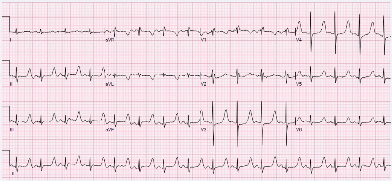
- ECG: prolonged QT or ST segment, arrhythmia
Outside hemorrhage, you treat the symptoms or the ECG change. A number alone is not the trigger. Correct magnesium when it is low.
High (above 1.32 mmol/L)
A high iCa usually traces back to one of three things: hypercalcemia (see the Calcium page), an acidotic sample, or calcium given intravenously.
| Mechanism | Common causes | Bedside clue |
|---|---|---|
| Acidosis | Severe or lactic acidosis | Low pH with high iCa; iCa typically falls after the acidosis is corrected |
| Hypercalcemia | Malignancy, hyperparathyroidism, other causes | High total calcium with high iCa; see the Calcium page |
| Calcium excess | Overdose of IV calcium or calcium supplements | Recent calcium dose. The label warns that hypercalcemia from overdose raises the risk of digoxin toxicity |
Interpretation & Bedside Approach
Step-by-step approach
- Is it real? Check the sample: heparin type, air, hemolysis and delay. Liquid heparin and hemolysis lower the result.
- Read the pH from the same sample. A 0.1 unit rise in pH lowers iCa by about 0.05 mmol/L, and acidosis raises it. Repeat iCa after the pH is corrected.
- How sick is the patient? Tetany, seizure, laryngospasm, confusion or ECG change means treat now.
- Acute or changing? During massive transfusion iCa moves with each unit. Trend it with every blood gas: a trend beats a single value.
- Classify with the thresholds: below 1.1 mmol/L is mild, below 0.9 mmol/L needs prompt correction, and below 0.8 mmol/L carries dysrhythmia risk (2023 European guideline and the trauma meta-analysis definitions).
- Escalate or hand off. Send the last iCa, its time, the pH, the calcium given and the blood product volume to the receiving team.
Pattern recognition
| Pattern | Suggests |
|---|---|
| Low iCa during massive transfusion, normal pH | Citrate chelation: give calcium chloride 1 g and recheck |
| Low iCa with high pH | Alkalosis (hyperventilation, bicarbonate): correct the pH and recheck |
| Low iCa with liver failure or shock | Slow citrate clearance: calcium chloride is preferred |
| High iCa with low pH | Acidosis raising iCa: treat the acidosis and expect iCa to fall after resuscitation |
ECG clue
Hypocalcemia stretches the ST segment and the QT interval. The ECG shows that effect, but it does not measure iCa, so check the iCa. In Figure 1, read only the long ST segment and the QT interval. The rest of the tracing is not part of the calcium pattern.
Management Priorities
Emergency: Low ionized calcium during massive transfusion (iCa below 0.9 mmol/L, or hypocalcemic symptoms or arrhythmia)
- Monitor and draw iCa: continuous ECG; iCa from the blood gas with each MTP sample, and again after each calcium dose.
- Give calcium chloride first: 1 g (10 mL of 10%) IV, slowly, through a central or deep vein per the label. Calcium gluconate is the alternative. The 2026 Canadian consensus lists 1 g chloride or 3 g gluconate as alternative doses; both give about 270 mg elemental calcium (chloride 270 mg per 10 mL, gluconate 90 mg per 10 mL).
- Keep the rate within the label: maximum 1 mL/min (100 mg/min) per the US label, so 1 g takes at least 10 minutes. Stop for pain, hypotension or bradycardia. Some resuscitation protocols give calcium faster; that is outside the label and should be decided with medical direction.
- Repeat by level, not by the clock: aim for iCa within 1.1–1.3 mmol/L. Repeat doses guided by iCa and the ECG, and hold further IV calcium while iCa is above range.
No central or deep line: Calcium chloride's label points to a central or deep vein. When neither is in place, many protocols accept a large, well-functioning proximal peripheral IV, watched closely at the site. Follow local protocol and medical direction. Calcium gluconate is labeled for a secure IV line and is often picked for a peripheral IV. Keep it at or under 200 mg/min (see the table).
Medications
| Drug | Adult dose | Pediatric dose (max) | Onset / key notes |
|---|---|---|---|
| Calcium chloride 10% | 1 g (10 mL) IV, at most 1 mL/min, central or deep vein per the label; repeat guided by iCa and ECG per protocol; recheck for overshoot | 20 mg/kg (0.2 mL/kg) IV/IO, usually max 1 g per dose (protocol-based; see the calcium chloride page). The US label's pediatric statement is 2.7–5 mg/kg for acute symptomatic hypocalcemia, so the protocol figure is higher; follow local protocol and pharmacist or medical direction | Holds 270 mg elemental calcium per 10 mL; preferred when the liver is impaired; avoid with digoxin |
| Calcium gluconate 10% | 3 g (30 mL of 10%) IV, diluted to 10–50 mg/mL and given no faster than 200 mg/min (at least 15 min); repeat guided by iCa and ECG | Protocol-based: see the calcium gluconate page. Label maximum rate 100 mg/min in pediatric patients | Holds 90 mg elemental calcium per 10 mL, about one-third of the chloride; slower in liver dysfunction. Contraindicated in neonates up to 28 days on ceftriaxone (label) |
Other indications for IV calcium
- Hyperkalemia with ECG changes (see the calcium gluconate and calcium chloride pages)
- Calcium channel blocker or beta-blocker toxicity, as an adjunct to standard toxicology care
- Hypermagnesemia with ECG changes or hypotension
- Symptomatic hypocalcemia: tetany, seizure or arrhythmia
High iCa (above range)
- Confirm: repeat from a fresh, air-free sample; check total calcium with albumin, PTH and the pH per the Calcium page.
- Stop: hold further IV calcium and calcium or vitamin D supplements. After calcium dosing, a high value means stop and recheck.
- Assess: ECG for a short QT or arrhythmia; ask about vomiting, polyuria, confusion and weakness.
- Treat by severity: isotonic saline for volume depletion; calcitonin, bisphosphonate or dialysis for severe or symptomatic hypercalcemia, under specialist or ICU direction. Use loop diuretics cautiously, after volume repletion.
- If on digoxin: hypercalcemia raises digoxin toxicity risk; monitor the ECG and calcium closely.
Definitive care & disposition
- Hemorrhage control and transfusion come first: no trial has shown that preventing or treating low iCa lowers mortality in critical bleeding (2023 European guideline).
- Admission: admit patients who need a calcium infusion or repeated dosing, and trend iCa with each blood gas.
Doses are typical references. Follow local protocols, medical direction and institutional policy.
Field & Transport Considerations
Point-of-care testing
Blood-gas cartridges and point-of-care analyzers give iCa from whole blood, usually alongside the pH, and they do it within minutes. Most blood-gas analyzers carry iCa in their panel. Run it promptly from a balanced-heparin syringe, since heparin binds calcium. Take the iCa and the pH together, from one sample.
Before and during interfacility transport
| Setting | Consideration |
|---|---|
| Blood products running | Check iCa with each blood gas and before departure when available. The 2026 Canadian prehospital consensus recommends routine calcium at intervals, considered at minimum after the second unit |
| No point-of-care iCa | Give calcium on the protocol schedule (chloride 1 g or gluconate 3 g) and target a measured value once one is available |
| Digoxin on the medication list | Avoid calcium chloride. If it is essential, give it slowly with continuous ECG monitoring |
| Calcium infusion en route | Use a pump. The label directs a central or deep vein; where only a peripheral IV is available, follow protocol and medical direction, check the site often and stop at any sign of infiltration. Record the last iCa and its time |
Scope: IV calcium (Paramedic/RN/CCT) per local protocol and medical direction.
Special Populations
| Population | What changes | Clinical implication |
|---|---|---|
| Neonates & infants | Age-specific range; calcium chloride and calcium gluconate are both contraindicated with ceftriaxone up to 28 days of age | Use the laboratory's neonatal range. Do not give any IV calcium product to a neonate who is receiving or will receive ceftriaxone, even through separate lines or at different times |
| Children & adolescents | The chloride label gives a pediatric dose of 2.7–5 mg/kg; no pediatric iCa range is verified here | Dose by protocol and weight (max per the calcium chloride page); use the pediatric range on the report |
| Pregnancy | No pregnancy-specific iCa range verified | Use the laboratory range and the obstetric hemorrhage protocol; check iCa during transfusion |
| Older adults | No age-specific limit verified | Interpret as for adults: treat the trend and symptoms, not the age |
| Kidney failure & CRRT | Citrate is used to anticoagulate CRRT, and its load adds to calcium handling | Check iCa per the CRRT protocol before and during therapy; see the CRRT page |
| Liver disease | Citrate is cleared more slowly | Ionized hypocalcemia is more likely with blood products; calcium chloride is preferred (2023 European guideline) |
Interfering Factors & Pitfalls
Collection & handling
| Factor | Effect | Why | Avoid / fix |
|---|---|---|---|
| Liquid (therapeutic) heparin | ↓ iCa (False −) | Heparin binds calcium; liquid heparin also dilutes the sample | Use a balanced-heparin blood-gas syringe |
| Hemolysis | ↓ iCa (False −) | Massive hemolysis dilutes analytes inside cells, including calcium | Redraw and note the hemolysis |
| Air exposure | ↓ iCa (False −) | Loss of CO₂ raises pH, which increases calcium binding to albumin | Expel air, cap the syringe and analyze promptly |
| Delay before analysis (unseparated sample, room temperature) | ↑ iCa (False +) | Ongoing glycolysis produces lactate, so pH falls and iCa reads higher | Analyze promptly, within the laboratory's time limit |
Medications & products
| Factor | Effect | Why | Avoid / fix |
|---|---|---|---|
| Citrate in blood products | ↓ iCa (true) | Citrate chelates calcium | Trend iCa with transfusion; use calcium chloride when the liver is impaired |
| Digoxin | Arrhythmia risk with calcium | Hypercalcemia from calcium overdose raises digoxin toxicity risk (label) | Avoid calcium chloride unless essential; monitor ECG and calcium |
| Bicarbonate or hyperventilation | ↓ iCa (true) | Higher pH increases calcium binding to albumin | Recheck iCa after alkalinizing or changing ventilation |
Physiologic / biologic
| Factor | Effect | Why | Avoid / fix |
|---|---|---|---|
| Acidosis | ↑ iCa | Hydrogen ions displace calcium from albumin binding sites | Read iCa with the pH; repeat after resuscitation |
| Alkalosis | ↓ iCa | About 0.05 mmol/L fall per 0.1 unit rise in pH | Read iCa with the pH |
Common misinterpretations
- Chasing a low total calcium when the measured iCa is normal.
- Assuming a normal routine coagulation test rules out a calcium-related clotting defect.
- Swapping calcium chloride and calcium gluconate as if they were the same: chloride holds about three times the elemental calcium per mL.
- Assuming correcting iCa lowers mortality: no trial has shown that.
Physiology & Test Basics
What it measures
This page is about ionized (free) calcium, the fraction that is physiologically active. We measure it in whole blood on blood-gas and point-of-care analyzers. Total calcium (serum chemistry, see the Calcium page) is the ionized fraction plus albumin-bound and complexed calcium. Nerves, muscle, the heart and clotting respond only to the ionized fraction.
Physiology & regulation
- About half of total serum calcium is ionized. About 40% sits on protein, mainly albumin, and the rest is bound to acids (DailyMed label).
- Fibrin polymerization and platelet function both need ionized calcium. Low levels impair clot formation (2023 European guideline).
- Low iCa also gets in the way of cardiac contractility and systemic vascular resistance (2023 European guideline).
- iCa depends on pH. A 0.1 unit rise in pH lowers it by about 0.05 mmol/L.
- Citrate in transfused blood keeps binding calcium until the liver clears it.
How it is measured
- Specimen: whole blood in a balanced-heparin blood-gas syringe, free of air, analyzed promptly. Liquid heparin both binds calcium and dilutes the sample.
- Where: blood-gas and point-of-care analyzers give you iCa within minutes, along with the pH from the same sample.
Why it matters
Low iCa weakens clotting, contractility and vascular tone. Standard tests will not reveal that, since the sample is citrated and recalcified before analysis (2023 European guideline). Measure the iCa directly.
Clinical Pearls
References
- Rossaint, R., Afshari, A., Bouillon, B., et al. (2023). The European guideline on management of major bleeding and coagulopathy following trauma: sixth edition. Critical Care, 27, 80. https://doi.org/10.1186/s13054-023-04327-7
- Peddle, M., Greene, A., Trojanowski, J., et al. (2026). Best practices on blood and blood products for a prehospital hemorrhage protocol: consensus from the 2025 Canadian prehospital transfusion summit. Scandinavian Journal of Trauma, Resuscitation and Emergency Medicine, 34 (Suppl 1), 100. https://doi.org/10.1186/s13049-026-01630-4
- Palitnonkiat, V., Thampongsa, T., Sirikun, J., & Owattanapanich, N. (2025). Association between hypocalcemia and mortality in trauma patients: a systematic review and meta-analysis. Trauma Surgery & Acute Care Open, 10(4), e001800. https://doi.org/10.1136/tsaco-2025-001800
- Dupuy, C., Martinez, T., Duranteau, O., et al. (2025). Comparison of the lethal triad and the lethal diamond in severe trauma patients: a multicenter cohort. World Journal of Emergency Surgery. https://doi.org/10.1186/s13017-024-00572-5
- Rajesh, A., Barry, L., Limon, D., et al. (2026). Aggressive calcium chloride dosing reduces early mortality in trauma patients receiving whole blood resuscitation. Journal of Trauma and Acute Care Surgery, 101, 57–64. https://doi.org/10.1097/ta.0000000000005009
- Hewgley, W. P., Griffin, R. L., Baird, E., et al. (2026). Early calcium replacement after minimal blood transfusion is associated with adverse outcomes. European Journal of Trauma and Emergency Surgery, 52. https://doi.org/10.1007/s00068-026-03282-6
- PAI Pharma. (2026). Calcium chloride injection, USP 10%: prescribing information. DailyMed, U.S. National Library of Medicine. https://dailymed.nlm.nih.gov/dailymed/drugInfo.cfm?setid=d5d9c301-599e-47bb-ba9b-c0065c08dfb4
- Baird, G. (2013). Preanalytical considerations in blood gas analysis. Biochemia Medica, 23, 19–27. https://doi.org/10.11613/bm.2013.005
Medical Disclaimer
- For Educational Purposes Only: This content is intended for educational reference and should not be used for clinical decision-making.
- Not a Substitute for Professional Judgment: Always consult your local protocols, institutional guidelines, and supervising physicians.
- Verify Before Acting: Users are responsible for verifying information through authoritative sources before any clinical application.