

Bedside Snapshot
- Indications
- Manual CPR impractical or unsafe: moving ambulance, cath lab, stairs
- Prolonged resuscitation with falling compression quality
- Bridge to ECPR cannulation under local protocol
- Contraindications / Abort
- Not for routine use: ILCOR 2026 and current guidance advise against it
- Patient outside device size or chest limits: check the IFU
- Device fails or cannot be placed quickly: resume manual CPR at once
- Critical Steps
- Pause manual CPR once and keep the pause short
- Place backplate or band, center the pad, start the device
- Keep defibrillation and drugs on the ACLS schedule
- Confirm & Monitor
- Continuous EtCO₂ and a pulse at each rhythm check
- Pad or band position after every move
- Pause time recorded and kept within protocol
Key Numbers
Mechanical CPR keeps compressions going when manual CPR is impractical or unsafe. Every device deployment must keep compressions interrupted for as short a time as possible.
| Parameter | Value |
|---|---|
| Routine use | Not recommended: no proven benefit over high-quality manual CPR; AHA 2025 and the 2026 ILCOR review advise against routine use |
| When to use | Manual compressions are difficult or dangerous to deliver: moving ambulance, cath lab, confined space, prolonged resuscitation, bridge to ECPR |
| Pause goal | Deployment and removal inside pauses of about 10 s or less (protocol target, not an AHA number) |
| Staffing | Two trained staff for deployment (protocol-dependent) |
| Survival in trials | No proven benefit: LINC 4-hour survival 23.6% mechanical vs 23.7% manual; harm signals in Where It Fits |
| Trial population | Adults only; trials excluded known pregnancy, traumatic arrest, under 18 years and bodies too large or small for the device |
| Pediatric | Not established: use manual CPR per pediatric protocol |
| Epinephrine | 1 mg IV/IO every 3–5 min: device use does not change drug timing |
| Amiodarone | 300 mg, then 150 mg for shock-refractory VF/pVT (per protocol) |
| Lidocaine (alternative) | 1–1.5 mg/kg IV/IO, then 0.5–0.75 mg/kg every 5–10 min Max 3 mg/kg; per protocol |
Overview & Principles
A mechanical device does the compressing. That means compressions keep going when a rescuer cannot deliver them safely or consistently. Two device families are in use. The machine does not change ACLS: rhythm checks, shocks and drugs stay on the same timing.
How It Works
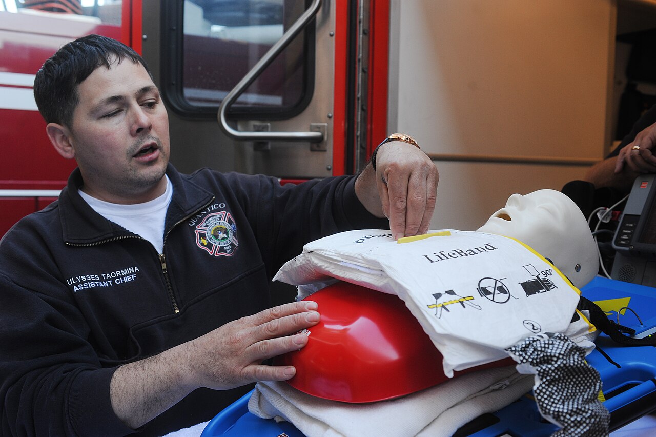
- Piston-type device: the pad sits on the lower sternum. A piston drives it down against a backplate slid under the patient.
- Band-type device: a load-distributing band wraps the chest and cinches down on a backboard. Compressions spread over the whole chest.
- Deployment: manual compressions stop for a moment while the backplate and device go on. Then the device starts.
- Verification: the device is a compression source and nothing more. Pulse, EtCO₂ and rhythm checks still tell you whether the patient is perfused.
| Device type | How it compresses | Practical note |
|---|---|---|
| Piston (e.g., LUCAS-type) | Pad on lower sternum, piston and backplate | Rapid to attach on a stretcher; pad position must be checked after every move |
| Band (e.g., AutoPulse-type) | Load-distributing chest band on a backboard | Band tension and fit must be checked; size and chest shape limits per IFU |
Where It Fits
Don't reach for it by default. AHA 2025 and the 2026 ILCOR systematic review both advise against routine use of mechanical CPR devices. Both still leave room for settings where high-quality manual compressions are difficult or dangerous for the rescuer. The condition: interruptions stay strictly limited during deployment and removal.
Look at the trial evidence and no benefit shows up. Taken overall, the three largest trials showed no benefit, or harm. PARAMEDIC (LUCAS-2) found 30-day survival of 6% vs 7%, and a worse adjusted 3-month neurologic outcome (aOR 0.72, 95% CI 0.52–0.99). ASPIRE, a band device, had fewer patients with a favorable neurologic outcome at discharge (RR 0.41, 95% CI 0.21–0.79). One trial suggests shockable-rhythm outcomes may have suffered from first shocks that came late, after early placement.
Indications & Contraindications
Indications
- Transport during arrest: the patient is moving or the ambulance is. Manual compressions turn unsafe or ineffective (ILCOR names transport as an example).
- Confined or hazardous spaces: stairs, elevators, small rooms or the cath lab. Anywhere a rescuer cannot compress safely.
- Prolonged resuscitation: rescuers tire, and compression quality falls with them.
- Bridge to ECPR: keeps compressions going while extracorporeal cannulation is set up. Follow the local ECPR protocol.
Absolute Contraindications
- None for a patient in cardiac arrest, provided a trained team can deploy the device without a long pause. The manufacturer IFU sets device size and chest limits.
Relative Contraindications & Cautions
- Patient too large or too small for the device: the LINC trial excluded them. Check the IFU before you choose a device.
- Suspected pregnancy: the trials excluded it. Follow the obstetric arrest pathway per local protocol.
- Traumatic arrest: the large trials excluded it. Apply the trauma arrest protocol first.
- ROSC or a conscious patient: skip the device when compressions are not needed.
- No trained operator: a badly deployed device means long pauses, and worse CPR than a practiced manual team gives.
Consider an Alternative
- High-quality manual CPR: rotate two rescuers about every 2 min, with a feedback device where you have one.
- Defibrillation first: for a shockable rhythm, see the defibrillation page.
Scope & Team Roles
| Provider Level | Typical Role | Notes |
|---|---|---|
| EMT | Assists | Helps position the backboard and hold manual compressions during deployment |
| AEMT | Assists | Per local protocol |
| Paramedic | Performs (protocol/credentialing required) | Device deployment often requires agency credentialing |
| RN (ED/ICU) | Assists | Medication and monitoring role during transport |
| Critical Care Transport (CFRN / CCP-C / FP-C) | Performs (protocol/credentialing required) | Common in transport; device use per program protocol |
| Physician / APP | Assists | Directs use through medical direction and ECPR criteria |
Team Roles During the Procedure
- Operator: places the pad or band and starts the device.
- Airway and ventilation: keeps the airway and breaths going during the pause.
- Medication clinician: keeps drug timing on the rhythm-check schedule.
- Monitor and recorder: times pauses and records EtCO₂, rhythm and device events.
Anatomy & Landmarks
| Site | Best For | Landmark | Angle/Depth | Avoid If |
|---|---|---|---|---|
| Piston pad | Stretcher and transport | Lower half of sternum | Per IFU | Chest injury over the sternum, or pad cannot be centered |
| Chest band | Backboard and transport | Band centered on the chest per IFU | Per IFU | Chest size outside the device range |
Piston Pad
- Expose the chest: remove clothing. Do not remove working defibrillation or pacing pads to place the device; if pads are not yet on, apply them in an anterolateral or anteroposterior position clear of the pad or band.
- Find the lower half of the sternum: keep the pad above the xiphoid.
- Place the backplate: slide it under the back during a pause, then attach the pad and piston.
- Confirm the pad is centered: check that it does not slip toward the abdomen or the upper chest.
Chest Band
- Place the backboard: the patient must be supine on a firm surface.
- Pass the band under the patient: lay it flat on the backboard and roll the patient gently.
- Secure the band: fasten it snug per IFU, with the chest centered in the band.
Equipment & Preparation
Equipment
- Mechanical CPR device: piston pad with backplate, or band with backboard; check battery charge and spare battery.
- Backboard: firm, flat surface for band devices and for stretcher transport.
- Defibrillator: pads applied before deployment, in a position clear of the compression pad or band. Do not move working pads to make room.
- Waveform capnography: continuous EtCO₂ for confirmation during the arrest.
- Backup: a trained manual compressor and a second device or manual kit if the first fails.
Sizing
Every device has size and chest limits, and the IFU publishes them. Trial patients who were too big or too small for the device were excluded. For a child or a very large patient, read the IFU before you choose.
Patient Positioning & Preparation
Flat on the back, on a firm surface. Put the defibrillator pads on and set the airway plan before the device goes on. That keeps those jobs out of the pause.

Pre-Procedure Checklist
- Confirm the indication: manual CPR is impractical or unsafe
- Battery charged; spare battery in the bag
- Device size checked against patient weight and chest size
- Backboard or firm surface under the patient
- Defibrillator pads applied before deployment
- Capnography connected and reading
- Two trained staff assigned to deployment
- Manual compressor ready as backup
- Pause timer started at the first interruption
Medications & Settings
These are typical adult and pediatric references. Check them against local protocol and current drug labeling.
| Drug | Adult Dose | Pediatric Dose | Onset / Duration | Notes |
|---|---|---|---|---|
| Epinephrine | 1 mg IV/IO every 3–5 min No fixed max per cardiac arrest | 0.01 mg/kg IV/IO every 3–5 min (max 1 mg) | IV/IO: rapid onset; short duration, so redose on the 3–5 min schedule | High-alert Drug timing does not change with the device. Give on the rhythm-check schedule. |
| Amiodarone | 300 mg IV/IO, then 150 mg for shock-refractory VF/pVT Per protocol | Per pediatric protocol | IV: onset within minutes | High-alert IV antiarrhythmic: watch BP and rhythm after each dose. |
| Lidocaine | 1–1.5 mg/kg IV/IO, then 0.5–0.75 mg/kg every 5–10 min Max 3 mg/kg; per protocol | Per pediatric protocol | IV/IO: rapid onset | High-alert Alternative to amiodarone for shock-refractory VF/pVT. Watch rhythm, BP and neurologic signs (CNS toxicity). |
Each device presets its own rate and depth. Check the manufacturer IFU for your unit, then compare the output with the manual target of 100–120/min and 5–6 cm. In the field, pause duration is the setting that matters most.
Step-by-Step Procedure
Phase 1: Prepare
- Confirm the need: manual CPR is impractical or unsafe for this patient and setting.
- Pick the device: confirm size, battery and backboard before the pause.
- Assign roles: two trained staff deploy the device; one keeps the airway and ventilation going.
Phase 2: Deploy
- Pause manual CPR: start the pause timer at the first interruption.
- Place the backplate or band: move the patient only as far as needed, with the backboard under them.
- Attach and start the device: confirm the pad or band is centered before the first compression.
- Confirm the device is compressing: the chest visibly compresses and recoils fully, and an EtCO₂ waveform is present. A low or falling EtCO₂ means check position and device output.
- Resume the ACLS routine: rhythm checks, shocks and drugs continue on the same schedule.
Phase 3: Confirm & Transport
- Check the device at each move: pad or band position and tension are rechecked after every move.
- Remove the device only at a pause: manual compressions resume immediately after removal.
Alternative Technique: Manual CPR Backup
The device fails, will not go on quickly, or the patient cannot be secured to the backboard? Return to manual CPR at once. Do not spend the pause troubleshooting the device.
Confirmation & Monitoring
Primary Confirmation
A running device is not proof of effective compressions. Check continuous EtCO₂ and the pulse at each rhythm check, and confirm perfusion with both. EtCO₂ is a trend of perfusion quality, not a target. Low or falling? Check position, rhythm and device output. A sudden, sustained rise can signal ROSC.
Secondary Confirmation
- Pad or band position: centered and secure, without slipping toward the abdomen
- Compression depth: the device reports depth per the IFU. Still, watch the chest move.
- Pause duration: recorded by the monitor and kept within the protocol
| Finding | Success | Failure / Malposition |
|---|---|---|
| EtCO₂ | Waveform present; a sudden, sustained rise can signal ROSC | Low or falling EtCO₂: check position, rhythm and device output |
| Pulse | Palpable pulse with ROSC | No pulse at a rhythm check: continue CPR per protocol |
| Pad / band | Centered on the sternum or chest | Off-center or slipping: stop, reposition, resume |
| Pauses | Within the protocol target | Repeated long pauses: identify the cause and shorten |
Ongoing Monitoring
- Every 2 min: rhythm check with EtCO₂ and pulse.
- Pulse checks: palpation is unreliable while the device compresses. Check the pulse in the planned rhythm-check pause, or use an arterial line or ultrasound where available.
- After each move: device position, tension and battery.
- After ROSC: remove the device per protocol and reassess perfusion.
Troubleshooting
| Problem | Likely Cause | Fix |
|---|---|---|
| Pad slips off-centre | Patient moved or pad not seated | Pause, reposition, restart per IFU |
| Band loosens | Patient moved on the backboard | Pause, retension, check the chest is centered |
| Low EtCO₂ with device running | Poor compression, pad off-center or low perfusion | Check position; pause briefly; resume manual CPR if needed |
| Device alarm or stop | Battery, fault or mis-seated component | Return to manual CPR at once; fix or replace the device |
| Long pause during deployment | Too few staff or equipment not ready | Stop, resume manual CPR, rehearse the deployment sequence |
| Rib or chest injury noted | Device or patient position | Reposition; document; reassess injuries |
If It Still Fails
- Resume manual CPR: at once, with a second rescuer rotating.
- Defibrillate on time: per the ACLS algorithm; see the defibrillation page.
- Escalate: to ECPR criteria under local protocol, if the team and system support it.
Complications
| Complication | Recognition | Prevention | Management |
|---|---|---|---|
| Immediate: chest bruising | Bruising under the pad or band after the arrest | Correct pad position; keep the device centered | Document; reassess position and injuries |
| Immediate: chest laceration or blood from mouth | Skin wound or blood at the airway after deployment | Check skin and airway before each move | Control bleeding, suction, reassess airway |
| Delayed: rib or sternal fracture | Chest pain, crepitus or pain after ROSC | Center the pad or band per IFU and recheck position after each move | Reassess and document; treat per protocol |
| Delayed: visceral or thoracic injury (liver or spleen laceration, pneumothorax) | Abdominal pain or distension, hypotension or respiratory compromise after ROSC | Center the pad or band per IFU; reassess position after every move | Image and treat per local protocol; involve surgical or thoracic team early |
| Rare: device malfunction | Alarm, stop or uneven compressions | Battery check; spare device or manual backup | Return to manual CPR at once |
| Rare: prolonged pauses | Pauses beyond the protocol target | Rehearsed deployment with two trained staff | Resume manual CPR; review the event |
PARAMEDIC recorded no serious adverse events. It did record seven clinical adverse events in the device group (chest bruising, chest lacerations and blood in the mouth), and none in the manual group. ILCOR 2026 found no significant difference in serious resuscitation-related or CPR-related injuries across the trials that reported them. Certainty is low. A 2023 retrospective cohort of 335 patients found abdominal injuries after skeletal and thoracic injuries. It also found more CPR-related injuries in device-assisted patients (aOR 6.2, 95% CI 2.5–15.4). It is observational, so treat it as a signal: check position and reassess.
Special Populations
Pediatrics
- No pediatric evidence: the large trials enrolled adults only. Children get manual CPR per the pediatric protocol. Do not put an adult device on a child.
- Sizing and dosing: see Equipment Sizing, Weight-Based Dosing and Pediatric Cardiac Arrest.
- Vascular access: see Pediatric Vascular Access; the arrest drugs follow the pediatric algorithm.
Pregnancy
- Trial exclusion: the trials left out known pregnancy.
- Pathway: follow the obstetric arrest pathway per local protocol.
Trauma / C-spine
- Traumatic arrest: the large trials left it out. Apply the trauma arrest protocol first.
- Spinal care: maintain alignment during deployment and transport per local protocol.
Physiologically Difficult
- Very large or very small bodies: several trials had them outside the device range. Check the IFU before deployment.
Post-Procedure Care & Transport
Immediate Aftercare
- After ROSC: remove the device at a pause and reassess perfusion and skin.
- After termination: record the device and time; follow local termination rules.
- Post-ROSC care: see the post-cardiac arrest care page.
Ongoing Maintenance
- Battery: keep a charged spare on the unit.
- Skin: check under the pad or band at each move.
Transport Considerations
- Securing: the backboard and device are secured before the vehicle moves.
- Reassessment: check position and tension after every move, turn or lift.
- Vibration and noise: rely on EtCO₂ and the monitor when the device or ambulance is loud.
- Safety: the device is an alternative to unsafe manual CPR, not an excuse to ignore scene safety.
Document & Hand Off
- Time of arrest, time of device start, and total pause time
- Device type, size and pad or band position
- Confirmation method: EtCO₂ and pulse readings
- Medications given and times
- Complications, including skin or chest injuries
- Response: ROSC time or termination decision
Clinical Pearls & Pitfalls
Pearls
- Deploy during a planned pause: stop, place, start, then resume the ACLS schedule.
- Pre-place pads: defibrillator pads go on before the device. The pause then never gets spent on them.
- Practice the sequence: two staff deploying the device should rehearse it on a manikin.
- Trust the numbers: EtCO₂ and pulse show perfusion. The device display does not.
Common Pitfalls
- Long deployment → rehearse: practice the deployment until it fits the protocol pause target.
- Device replaces manual skills → keep manual CPR first: manual CPR remains the default.
- Device running with no pulse → check perfusion: confirm with EtCO₂ and pulse, then decide.
Education & Skill Maintenance
Skill Maintenance
- Simulation: practice deployment at least every quarter on a manikin.
- Checklist: use the pre-procedure checklist during each drill.
- Debriefing: review pause times and device placement after each drill.
- Familiarization: learn the battery and fault indicators on your unit.
References
- Kleinman ME, Buick JE, Huber N, et al. Part 7: Adult Basic Life Support: 2025 American Heart Association Guidelines for Cardiopulmonary Resuscitation and Emergency Cardiovascular Care. Circulation. 2025;152(16_suppl_2):S448–S478. https://doi.org/10.1161/CIR.0000000000001369
- Wigginton JG, Agarwal S, Bartos JA, et al. Part 9: Adult Advanced Life Support: 2025 American Heart Association Guidelines for Cardiopulmonary Resuscitation and Emergency Cardiovascular Care. Circulation. 2025;152(16_suppl_2):S538–S577. https://doi.org/10.1161/CIR.0000000000001376
- Pocock H, Nicholson TC, Szarpak L, et al. Mechanical versus manual chest compression in cardiac arrest: a systematic review (ILCOR Advanced Life Support Task Force). Resuscitation Plus. 2026;29:101311. https://doi.org/10.1016/j.resplu.2026.101311
- Rubertsson S, Lindgren E, Smekal D, et al. Mechanical chest compressions and simultaneous defibrillation vs conventional cardiopulmonary resuscitation in out-of-hospital cardiac arrest: the LINC randomized trial (landmark trial). JAMA. 2014;311(1):53–61. https://doi.org/10.1001/jama.2013.282538
- Perkins GD, Lall R, Quinn T, et al. Mechanical versus manual chest compression for out-of-hospital cardiac arrest (PARAMEDIC): a pragmatic, cluster randomised controlled trial (landmark trial). Lancet. 2015;385(9972):947–955. https://doi.org/10.1016/S0140-6736(14)61886-9
- Hallstrom A, Rea TD, Sayre MR, et al. Manual chest compression vs use of an automated chest compression device during resuscitation following out-of-hospital cardiac arrest: a randomized trial. JAMA. 2006;295(22):2620–2628. https://doi.org/10.1001/jama.295.22.2620
- Preda T, Nafi M, Villa M, Cassina T. Traumatic injuries after manual and automatic mechanical compression during cardiopulmonary resuscitation, a retrospective cohort study. Resuscitation Plus. 2023;article 100465. https://doi.org/10.1016/j.resplu.2023.100465
- Merino JL, Tamargo J, Blomström-Lundqvist C, et al. Practical compendium of antiarrhythmic drugs: a clinical consensus statement of the European Heart Rhythm Association of the European Society of Cardiology. Europace. 2025. https://doi.org/10.1093/europace/euaf076
Medical Disclaimer
- For Educational Purposes Only: This content is intended for educational reference and should not be used for clinical decision-making.
- Not a Substitute for Professional Judgment: Always consult your local protocols, institutional guidelines, and supervising physicians.
- Verify Before Acting: Users are responsible for verifying information through authoritative sources before any clinical application.