

Bedside Snapshot
- Recognize
- Heart rate typically <50/min with a slow rhythm that causes hypotension, altered mental status, ischemic chest pain, acute heart failure or shock
- Red Flags
- Wide-QRS escape at 20–40/min; Mobitz II or complete block with symptoms; pauses; SBP falling; new confusion; chest pain
- First Actions
- Check pulse and perfusion; oxygen and monitor; 12-lead ECG; IV/IO access; atropine 1 mg; pads on early for pacing
- Key Doses
- Atropine 1 mg IV/IO, repeat every 3–5 min (max 3 mg); if no response, pace, or dopamine 5–20 mcg/kg/min or epinephrine 2–10 mcg/min. Child: epinephrine 0.01 mg/kg IV/IO with CPR if HR under 60 and poor perfusion
- Don't Miss
- Atropine is less likely to work in Mobitz II and complete block and can worsen it: pads on at once, and do not wait through a full atropine course before pacing. Asystole or PEA is not bradycardia: start CPR
- Escalate & Transport
- Escalate to transcutaneous pacing when atropine fails and to expert consultation for transvenous pacing; transport to a monitored ED or ICU bed with pacing available
Definition & Pathophysiology
Bradycardia is a heart rate below 50/min that is causing cardiopulmonary compromise. The number alone is not the trigger. The AHA 2025 adult algorithm asks whether the slow rhythm is hurting the patient. Slow rhythms come from the sinus node, or from conduction failure at the AV node or below it (the His-Purkinje system).
- Nodal (AV node) block: Narrow QRS escape at about 40–60/min. Often transient, and more likely to respond to atropine.
- Infranodal block: Wide QRS escape at about 20–40/min. Often persistent, less reliable, and more likely to asystole. Atropine is often ineffective, and it can worsen the block by speeding the atrial rate (see Atropine)
- Sinus bradycardia: Common with vagal tone, sleep, athletic training and beta-blockers. Benign when the patient is well.
- Reversible causes: hypoxia, hypovolemia, acidosis, hypo- or hyperkalemia, hypoglycemia, hypothermia, raised intracranial pressure (Cushing response), spinal cord injury with neurogenic shock, toxins (β-blockers, calcium channel blockers, digoxin, opioid or sedative effect, cholinesterase inhibitors or organophosphates), tamponade, tension pneumothorax and coronary or pulmonary thrombosis (the AHA H's and T's)
- Ischemic causes: Inferior MI often causes transient AV-node block. Anterior MI with septal involvement often causes infranodal block, and that carries a higher risk.
Recognition & Red Flags
Symptomatic Bradycardia
- Slow pulse with poor perfusion: hypotension, altered mental status, ischemic chest pain, acute heart failure or shock
- Rhythm pattern: sinus bradycardia, AV block (second- or third-degree) or a junctional or ventricular escape rhythm
- Onset: Sudden with an acute MI, drug toxicity or electrolyte change. Gradual in degenerative disease.
Stable or Asymptomatic Bradycardia
Normal perfusion and mental status don't end the work-up. Monitor the patient, get a 12-lead ECG, search for reversible causes and review the medications. Treat it as a deteriorating patient if the rate falls or the escape rhythm widens.
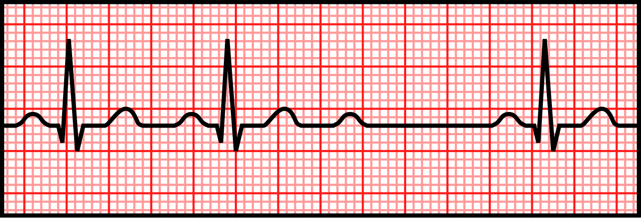
Red Flags
- Heart rate below 50/min with hypotension, altered mental status, ischemic chest pain, acute heart failure or shock
- Escape rhythm below 40/min, or a wide QRS escape
- Pauses between QRS complexes with no P wave, or dropped beats in a fixed pattern (Mobitz II)
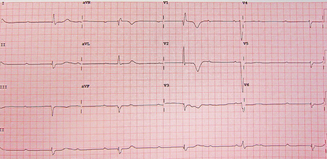
- AV dissociation, meaning P waves and QRS complexes beat independently (complete block)
Assessment & Diagnostics
Perfusion comes first. Then identify the block site. The treatment decision turns on symptoms, and on whether the QRS is narrow or wide.
| Check | Why | Act If |
|---|---|---|
| Pulse and perfusion | Decides between the bradycardia pathway and the arrest pathway | No pulse: start CPR and the ACLS arrest algorithm. Pulse with hypotension or altered mental status: treat as symptomatic |
| 12-lead ECG and rhythm strip | Block site and ischemia; a longer strip shows whether PR lengthens | Wide QRS escape or Mobitz II: atropine less likely to work; get pads on early. Ischemic changes: activate the STEMI pathway |
| SpO₂, ventilation and EtCO₂ | Hypoxia is a reversible cause and the first target in children | Hypoxia: oxygen and airway support before more drugs; in children, see the pediatric box under ALS Management |
| Potassium, magnesium and glucose | Abnormal result or ECG change: treat per protocol and notify ED early | |
| Medication and toxin history | β-blockers, calcium channel blockers, digoxin and cholinesterase inhibitors cause bradycardia | Suspected toxin: toxin-specific therapy early; call poison control or toxicology |
Use the QRS width to judge how likely atropine is to work. Get pads on early either way. A narrow QRS suggests a nodal site that may respond to atropine. A wide QRS suggests an infranodal site, and that site is unlikely to respond.
Differential & Mimics
- Sinus bradycardia: A regular sinus rhythm with normal PR and a narrow QRS. Benign in well patients. Treat when symptomatic.
- Sinus arrest or exit block: A pause with no P wave in front of it. The sinus node may still be firing.
- Mobitz I (Wenckebach): The PR lengthens across a group before a dropped QRS. Usually nodal, and it seldom needs pacing unless symptomatic.
- Junctional rhythm: Narrow QRS with a slow escape. Check for retrograde P waves.
- Atrial fibrillation with slow ventricular response: Irregular R-R with no P waves. Review the rate-slowing drugs, and check the ECG for conduction disease.
- Artifact or lead failure: A monitor that counts artifact as beats. Confirm with a 12-lead and a pulse check.
BLS Management
- Scene and patient safety: confirm a pulse and check breathing.
- Pulse absent: start CPR and the AED or defibrillator pathway; this is not a bradycardia case.
- Oxygen if hypoxic or breathing is inadequate; assist ventilation with a bag-valve-mask as trained.
- Position and reassess: keep the patient still, lie flat if hypotensive, and watch mental status and chest pain.
- Call ALS early and report the rhythm, rate, blood pressure, symptoms and time of onset.
BLS staff don't give atropine, and they don't start pacing. If the ALS unit is delayed, put the monitor and pads on, per local protocol.
ALS Management
The 2025 AHA adult bradycardia algorithm and Part 9 are the source for everything below.
- Atropine first (Paramedic/RN/CCT): A 1 mg IV/IO bolus, repeated every 3–5 min, tops out at 3 mg total. The 2025 AHA adult algorithm still starts with atropine when bradycardia causes compromise (Part 9, COR 2a, LOE B-NR). Don't shave the dose. Give the full 1 mg, because a smaller one can slow the rate first and speed it up only after. See Atropine.
- Atropine in infranodal block: Atropine still goes first on the algorithm. Will it work in Mobitz II, complete block or a wide escape? Less likely, and it can worsen the block. So the pads go on at the same time. Don't sit through a full atropine course before you pace.
- Pads and transcutaneous pacing (Paramedic/RN/CCT): Pads on early. Atropine fails? Pace (2025 AHA Part 9 algorithm). In unstable high-degree AV block with no IV or IO access, immediate pacing may be considered (COR 2b, LOE C-EO). The rate and output live on the pacing page. Give analgesia or sedation before the first spike. See Transcutaneous Pacing.
- Infusion as a bridge (Paramedic/RN/CCT): Dopamine runs at 5–20 mcg/kg/min IV, dosed by weight. Titrate it to heart rate and perfusion while you set up pacing or if pacing fails (2025 AHA Part 9, COR 2b, LOE C-LD). Use it when atropine is ineffective. It is off-label for bradycardia. See Dopamine.
- Epinephrine alternative: Run 2–10 mcg/min IV infusion at a fixed rate, not by weight (2025 AHA Part 9, COR 2b, LOE C-LD). Use it when atropine is ineffective. It is off-label for bradycardia. Titrate to response. See Epinephrine IV.
- Toxic causes: Start toxin-specific therapy early, and call poison control or toxicology (1-800-222-1222). For hypotension, vasopressors are recommended (2025 AHA Part 10: COR 1, LOE C-EO for β-blocker poisoning, COR 1, LOE C-LD for calcium channel blocker poisoning). High-dose insulin is recommended when β-blocker or calcium channel blocker poisoning brings hypotension that stays refractory to vasopressors (COR 1, LOE C-LD for β-blocker poisoning, and COR 1, LOE B-NR for calcium channel blocker poisoning). For β-blocker poisoning, a glucagon bolus then an infusion is reasonable when bradycardia or hypotension is symptomatic (COR 2a, LOE C-LD, see Glucagon). For digoxin toxicity, see Digoxin Immune Fab. Toxic bradycardia gets worse fast. If mental status declines, secure the airway early.
- Calcium: In calcium channel blocker poisoning, calcium is reasonable (2025 AHA Part 10, COR 2a, LOE C-EO). In β-blocker poisoning, it may be reasonable (COR 2b, LOE C-EO). See Calcium Chloride.
- Hyperkalemia: Treat per protocol. Calcium goes in first, to shield the myocardium while the potassium is shifted or removed. See Calcium Chloride.
- Reassess after every change: rate, rhythm, pulse at every paced beat, blood pressure and mental status.
ED & ICU Management
- Definitive pacing: Transvenous pacing for infranodal block, for symptomatic bradycardia that drugs don't fix, or for a pacing bridge that has to last longer than transcutaneous pacing can hold.
- Vasoactive support: Titrate the infusion to blood pressure and perfusion. Reassess at every change in pacing or drug dose.
- Diagnostics: Check troponin, electrolytes, a toxicology screen and an echocardiogram when the cause is not clear. See Conduction Blocks for ECG criteria.
- Ischemia: An acute MI with block is a cath lab emergency. Involve cardiology early.
- Consults: Cardiology or electrophysiology for a permanent pacing evaluation in Mobitz II and complete block. Toxicology for poisoning.
- Disposition: A monitored bed with pacing available. Don't discharge Mobitz II, complete block or a symptomatic bradycardia without a pacing plan.
Transport Considerations
- Destination: Go to the nearest ED that can provide pacing and cardiology. With an acute MI and block, that is the PCI-capable center.
- Pads on and pacing ready in transit. Check capture after every move, sedation change or position change.
- Infusion pump: Confirm the drug, the concentration and the weight-based rate (mcg/kg/min versus mcg/min) at the start, at every bag change and at handoff. Dopamine is a high-alert drug.
- Monitor continuously: Watch rhythm, pulse, blood pressure, SpO₂ and mental status. Check a pulse at each rhythm change.
- Handoff: time of onset, the rhythm strip, atropine doses and times, pacing thresholds and current infusion rate.
Special Populations
- Pediatrics: Most pediatric bradycardia is hypoxic or respiratory. Ventilate and oxygenate first. If HR is below 60/min with poor perfusion despite that support, start CPR and give epinephrine 0.01 mg/kg IV/IO (maximum 1 mg per dose), repeated every 3–5 min as in pediatric arrest (2025 AHA Part 8). Don't give atropine in place of CPR and epinephrine. Atropine 0.02 mg/kg (minimum 0.1 mg, maximum 0.5 mg, may be repeated once) is for increased vagal tone or primary AV block. See Pediatric Cardiac Arrest and Weight-Based Dosing.
- Pregnancy: Treat as you would a non-pregnant adult. The same drugs and pacing apply. During CPR, use manual left uterine displacement. Maternal hypotension threatens the fetus. Treat it early.
- Older adults: Older adults have lower cardiac reserve. A small change in rate can cause hypotension and confusion. Degenerative conduction disease is more common.
- Coronary artery disease or acute MI: The atropine label caps the total dose at 0.03–0.04 mg/kg (2–3 mg). The reason is atropine-induced tachycardia, which raises myocardial oxygen demand. At 70 kg, that is about 2.1–2.8 mg. The AHA algorithm allows up to 3 mg, so the cap is a protocol and medical-direction decision. See Atropine.
- Beta-blocker or calcium channel blocker use: Expect a poor response to atropine and a reduced response to epinephrine. Consider toxin-specific therapy early.
- Heart transplant: A denervated transplanted heart does not respond to atropine. Pace it, or use an infusion.
- Implanted pacemaker or ICD: Keep pads away from the generator, and follow the device plan.
- Renal and hepatic impairment: The atropine label gives no renal or hepatic dose adjustment. Reassess the rate after each dose.
Pitfalls & Safety Checks
- Error: Waiting through a full atropine course in Mobitz II or complete block. Pads go on at once. Be ready to pace if atropine is ineffective.
- Error: Underdosing atropine. A smaller dose can slow the rate before it speeds it up. Give the full dose and reassess.
- Error: Giving atropine to a hypoperfused child with HR below 60/min instead of starting CPR and epinephrine.
- Error: Pacing a patient in asystole or PEA. Pacing is not indicated here. Start CPR and the ACLS arrest algorithm.
- Error: Counting muscle twitching or a wide QRS as capture. Confirm capture with a pulse at every paced beat.
- Error: Giving dopamine as a push dose, or in the wrong concentration. Dopamine is an infusion only, and it is weight-based.
- Error: Confusing the bradycardia atropine cap with the organophosphate regimen. The 3 mg maximum applies to bradycardia. Organophosphate poisoning runs on a different schedule.
- Error: Pacing a conscious patient without sedation. Pacing hurts, and movement disrupts capture.
- Error: Missing a reversible cause such as hyperkalemia, hypoxia or a toxin. Check the cause at every reassessment.
Clinical Pearls
References
- Wigginton, J. G., Agarwal, S., Bartos, J. A., et al. (2025). Part 9: Adult advanced life support: 2025 American Heart Association Guidelines for Cardiopulmonary Resuscitation and Emergency Cardiovascular Care. Circulation, 152(16 suppl 2), S538–S577. https://doi.org/10.1161/CIR.0000000000001376
- Lasa, J. J., Dhillon, G. S., Duff, J. P., et al. (2025). Part 8: Pediatric advanced life support: 2025 American Heart Association and American Academy of Pediatrics Guidelines for Cardiopulmonary Resuscitation and Emergency Cardiovascular Care. Circulation, 152(16 suppl 2), S479–S537. https://doi.org/10.1161/CIR.0000000000001368
- Kusumoto, F. M., Schoenfeld, M. H., Barrett, C., et al. (2019). 2018 ACC/AHA/HRS guideline on the evaluation and management of patients with bradycardia and cardiac conduction delay. Circulation, 140(8), e382–e482. https://doi.org/10.1161/CIR.0000000000000628
- 2025 American Heart Association Guidelines for Cardiopulmonary Resuscitation and Emergency Cardiovascular Care, Part 10: Adult and Pediatric Special Circumstances of Resuscitation (2025). Recommendations for life-threatening β-blocker and calcium channel blocker poisoning (high-dose insulin, glucagon, calcium). https://cpr.heart.org/en/resuscitation-science/cpr-and-ecc-guidelines/adult-and-pediatric-special-circumstances-of-resuscitation
- Atropine sulfate injection [prescribing information]. Revised 9/2025. DailyMed, U.S. National Library of Medicine (2025). https://dailymed.nlm.nih.gov/dailymed/drugInfo.cfm?setid=ad8916e7-206e-409e-2582-30d072845dd4
- Epinephrine injection USP 1 mg/10 mL (0.1 mg/mL), for intravenous use [prescribing information]. DailyMed, U.S. National Library of Medicine (2024). https://dailymed.nlm.nih.gov/dailymed/drugInfo.cfm?setid=f2b5f15b-8541-466a-a27a-ee57bc4aac67
Medical Disclaimer
- For Educational Purposes Only: This content is intended for educational reference and should not be used for clinical decision-making.
- Not a Substitute for Professional Judgment: Always consult your local protocols, institutional guidelines, and supervising physicians.
- Verify Before Acting: Users are responsible for verifying information through authoritative sources before any clinical application.