

At a Glance
- Represents
- The impulse path: SA node → atria → AV node → His-Purkinje → ventricles. Each waveform marks one site.
- Normal (Adult)
- 60–100 bpm sinus, P before every QRS
- PR 120–200 ms
- QRS ≤110 ms
- Measure It
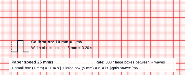
- Paper 25 mm/s (adult standard; read the label, since 50 mm/s is used in children): 1 small box = 0.04 s, 1 large box = 0.20 s. Gain 10 mm/mV; check the calibration pulse first.
- Abnormal → Think
- P waves without QRS, QRS without P waves, or a slow rate: name the site of failure before you name the rhythm.
- Red Flags
- Sinus pause (check pulse)
- Slow rhythm with no P waves
- Slow rate with hypotension or altered mental status
- Bedside Action
- Confirm P waves, rate and pulse, then treat per the current AHA bradycardia algorithm and local protocol.
Physiology & What It Represents
Every heartbeat starts in this system, and it reaches the ventricles in a set order. It starts at the sinoatrial (SA) node, the normal pacemaker in the right atrium. From there it spreads through the atrial muscle to the atrioventricular (AV) node. The AV node is slow on purpose. That physiological delay lets the atria empty before the ventricles contract.
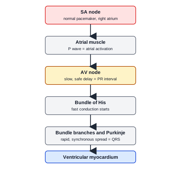
Past the AV node, the impulse moves fast. The bundle of His, the right and left bundle branches (the left one divides into fascicles) and the Purkinje network push it into the ventricular muscle in step. The chain runs by rank. The SA node normally sets the rate. Lower sites take over only when faster sites fail.
In most people, the right coronary artery feeds both the SA node and the AV node. An inferior infarction can therefore slow the rate or block conduction. See Conduction Blocks and STEMI Recognition.
On the ECG, each wave marks one site. The P wave is atrial activation. The PR interval is the AV node delay. The QRS is ventricular activation through the His-Purkinje system. When one wave changes, the change tells you where the problem is.
Normal Values & How to Measure
Check the paper speed and the gain before you measure anything. Every duration on the tracing depends on both.
| Parameter | Adult Normal | Pediatric Note | Where / How to Measure |
|---|---|---|---|
| Sinus rate | 60–100 bpm at rest | Age-dependent; see Pediatrics below | Count QRS in 6 s and multiply by 10, or divide 300 by the large boxes between R waves (regular rhythm) |
| PR interval | 120–200 ms (0.12–0.20 s; 3–5 small boxes) | Age-dependent | P onset to QRS onset in the lead with the clearest P (II) |
| QRS duration | ≤110 ms (≤2.75 small boxes); 111–119 ms is borderline; 120 ms or more is wide (3 small boxes or more) | Age-dependent | Start to end of the QRS in the widest lead |
| Paper speed | Standard 25 mm/s: 1 small box = 0.04 s; 1 large box = 0.20 s | Standard is 25 mm/s; 50 mm/s is often used for infants and children | Check the printed label on every tracing. At 50 mm/s: 1 small box = 0.02 s, 1 large box = 0.10 s; rate = 600 divided by large boxes between R waves |
| Gain | 10 mm/mV: 1 mV = 10 mm; 1 large box = 0.5 mV | Standard 10 mm/mV; some tracings use 5 or 20 mm/mV for large or small voltages | Check the calibration pulse (Figure 2) |
- Confirm the calibration: check the printed paper speed (adult standard 25 mm/s) and gain (10 mm/mV) before any measurement.
- Choose the lead: use lead II, or the lead with the clearest P waves.
- Measure PR: from the start of the P wave to the start of the QRS; convert small boxes at 0.04 s each.
- Measure QRS: in the widest lead, from its first deflection to its return to baseline.
- Calculate the rate: count R waves in 6 s (30 large boxes at 25 mm/s) and multiply by 10, or use 300 divided by large boxes between R waves for a regular rhythm (at 25 mm/s; at 50 mm/s use 600 divided by large boxes).
Sinoatrial Node Automaticity
The SA node fires without any outside prompt. Its cells creep toward threshold during diastole, then fire. That spontaneous creep sets the sinus rate. Autonomic input changes it.
ECG Criteria
- Sinus origin: a P wave precedes every QRS.
- Rate: 60–100 bpm at rest in adults.
What Sets the Rate
- Funny current (If): HCN4 channels carry the inward current behind that slow diastolic climb. Cyclic AMP acts on those channels.
- Sympathetic input: norepinephrine hits beta-adrenergic receptors. Cyclic AMP rises, and the node fires faster.
- Vagal tone: In humans, the resting heart rate reflects parasympathetic tone. That tone slows the sinus node.
Clinical Significance
- Normal rate change: The sinus rate rises and falls with autonomic tone. Read the rate alongside the rhythm and the patient.
- Sinus node dysfunction: When SA node firing fails, you get pauses or arrest. A lower site may take over, but at a slower rate.
Look-Alikes
- The full picture for sinus bradycardia, pauses and sinus arrhythmia is on Sinus Rhythms and ECG Rate.
AV Node Delay and the His-Purkinje System
Impulses normally reach the ventricles through the AV node, and the AV node is slow. That slowness shows up as the PR interval. Beyond it, the bundle of His, the bundle branches and the Purkinje fibers carry the signal fast.
ECG Criteria
- PR interval: 120–200 ms. A long PR points to delay at the AV node. A short PR points to a bypass pathway or a low atrial or junctional origin (see PR Interval and Junctional Rhythms).
- QRS width: Narrow when activation runs through the His-Purkinje system. 111–119 ms is borderline. 120 ms or more when activation starts in the ventricle or a bundle branch is blocked.
Escape Behavior
- Hierarchy: the SA node runs the ventricles at the fastest rate it can sustain.
- Escape: when the faster sites fail, a lower one takes over. Where the impulse starts sets both the rate and the QRS width (see Junctional Rhythms).
Clinical Significance
- Block location: a block at the AV node and a block below it give different escape rhythms and different pacing decisions. The detail is on Conduction Blocks.
- Purkinje junction: the Purkinje-ventricular junction gives reliable excitation. It can also set up conduction delay and ectopic activity.
Fast-Response and Slow-Response Action Potentials
Cardiac cells come in two types. Atrial and ventricular muscle upstroke fast in phase 0, then rest steady in phase 4. His-Purkinje cells also upstroke fast. They carry a latent phase 4 depolarization, slower than the rest, and that lets them take over as escape rhythms. SA and AV node cells are the other type. Their phase 4 is slow and spontaneous, and their upstroke is slower too because it depends on calcium currents.
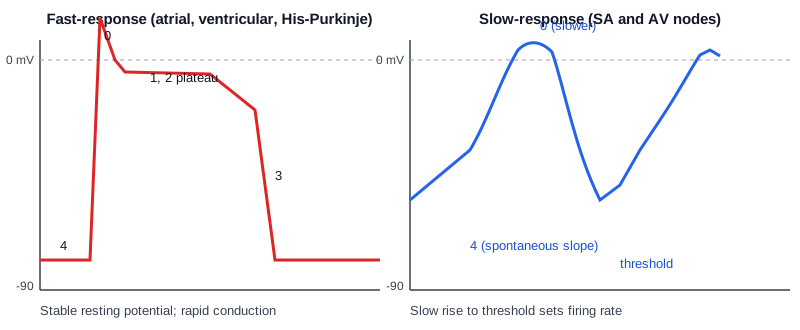
- Fast-response cells: atrial muscle, ventricular muscle and the His-Purkinje system. Conduction is rapid.
- Slow-response cells: SA and AV nodes. Conduction is slow, and they fire on their own, which sets the rate.
Why It Matters on the ECG
- Sinus rate: the spontaneous phase 4 slope of the SA node cells decides how often the node fires.
- PR interval: slow AV node conduction makes the PR interval. Fast His-Purkinje conduction keeps the QRS narrow.
Differential Diagnosis & Mimics
Sort the three patterns by where the impulse starts. Read the P wave and the QRS together. They show where the rhythm begins.
| Feature | Sinus (SA node) | Junctional (AV node) | Ventricular |
|---|---|---|---|
| P wave | Upright, before every QRS | Absent, inverted or after the QRS | Absent or dissociated |
| PR interval | 120–200 ms | Short or not measurable | Not measurable |
| QRS | Narrow | Usually narrow | Wide (120 ms or more) |
Artifact and Lead Misplacement
- Arm-lead reversal: lead I inverts and the axis flips. Check the axis before you call an ectopic or left-sided rhythm.
- Tremor or motion: baseline noise can hide small P waves or look like fibrillatory activity. Check a second lead.
- Vehicle vibration: on the move, artifact can look like a wide complex. Pause the motion or recheck when it is stable.
Bedside Management & Escalation
Conduction disease shifts your decisions on three points: the rate, how wide the QRS is, and whether the patient is still perfusing. No drug doses appear here. The linked pages and local protocols hold those.
- Sinus rhythm, adequate perfusion: monitor, obtain a 12-lead ECG where your scope allows, and treat the cause.
- Slow rate with poor perfusion (adults; for children see Pediatrics): treat it as symptomatic bradycardia. Follow the current AHA adult bradycardia algorithm: atropine first, then transcutaneous pacing or infusion per protocol (see Atropine and Transcutaneous Pacing). With an infranodal block (Mobitz II, or third-degree block with a wide QRS), atropine is unlikely to help and can worsen the block. After cardiac transplant it is ineffective, so pace early in those cases. Hunt for ischemia and the reversible causes: hypoxia, beta-blocker or calcium-channel blocker toxicity, hyperkalemia and hypothermia. (Paramedic/RN/CCT)
- Pause: check the pulse and perfusion first. Pulse present and symptomatic: manage as symptomatic bradycardia (see the bullet above). Pulseless, or unresponsive with no normal breathing: start CPR and the cardiac arrest algorithm, and notify online medical direction early. Pulse present and asymptomatic: monitor and look for reversible causes. Do not start CPR for a pause in a patient with a pulse.
Follow local protocols and medical direction.
Pediatric & Special Populations
Pediatrics
- Heart rate: normal rates change with age. Use the age table on Pediatric Vital Signs, not the adult 60–100 bpm.
- PR and QRS: both shift with age. Compare them to pediatric references, not adult cut-offs.
- Bradycardia with poor perfusion: in children it is usually hypoxic or ischemic. Support airway and breathing first. Then follow the AHA PALS bradycardia algorithm: start CPR if the heart rate is below 60 with poor perfusion despite oxygenation and ventilation. Don't run the adult bradycardia sequence unchanged.
- Doses: none on this page. Weight-based dosing lives on Weight-Based Dosing.
Other Populations
- Paced rhythms: a pacing spike with a wide QRS is the pacemaker at work, not native conduction. Check capture and the pacemaker type.
- Autonomic tone: a low resting rate may reflect high vagal tone, not disease. Judge it by the symptoms.
- Athletes: in well-trained athletes, a resting sinus rate below 60 and first-degree AV block can be normal. That usually comes with high vagal tone. Judge them by symptoms and the rest of the tracing.
- Older adults and medications: conduction disease gets more common with age. Beta-blockers, calcium-channel blockers, digoxin and antiarrhythmics slow the SA or AV node. Review them with any new bradycardia or block.
Monitoring & Transport Considerations
- Monitoring lead: lead II shows P waves best. Keep it on the monitor for rhythm checks.
- Artifact in transport: movement and vibration can bury P waves. Reposition the electrodes, or recheck with a 12-lead when stable.
- Calibration on handoff: write down the paper speed and gain on any printed tracing before anyone interprets it.
- Pacing check: after transcutaneous pacing, confirm mechanical capture with a femoral pulse, a pleth or SpO₂ waveform, or an arterial line. Muscle twitching can pass for a carotid pulse. The ECG alone does not confirm capture.
- Medications: when the rhythm slows or a block shows up, review the drugs that slow conduction: beta-blockers, calcium-channel blockers, digoxin.
Step-by-Step Approach & Pitfalls
- Confirm the calibration: paper speed and gain from the printed label (adult standard 25 mm/s and 10 mm/mV).
- Find the P waves: use lead II; check whether each QRS has a P before it.
- Measure the PR interval: 120–200 ms is normal.
- Measure the QRS: ≤110 ms is normal; 111–119 ms is borderline; 120 ms or more is wide.
- Count the rate: 6-second count times 10, or 300 divided by large boxes (at 25 mm/s; at 50 mm/s use 600 divided by large boxes).
- Name the origin and the site: sinus, junctional or ventricular; then the block, if any.
- Check perfusion and act: treat the patient per local protocol and medical direction.
Common Pitfalls
- Measuring with the wrong box size because nobody checked the paper speed.
- Calling a slow regular rhythm sinus bradycardia when there are no P waves.
- Measuring PR in a lead where you can't see the P wave.
- Reading amplitudes off a tracing whose gain you don't know.
ECG Examples
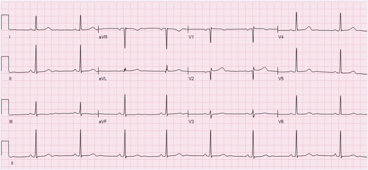
Here is the sinus origin this page describes. Every QRS has a P wave in front of it. The PR and QRS intervals are normal, and the rate is slow.
Clinical Pearls
References
- American Heart Association. Part 1: Executive Summary: 2025 American Heart Association Guidelines for Cardiopulmonary Resuscitation and Emergency Cardiovascular Care. Circulation. 2025;152:S284–S312. https://doi.org/10.1161/CIR.0000000000001372
- American College of Cardiology, American Heart Association, Heart Rhythm Society. 2018 ACC/AHA/HRS Guideline on the Evaluation and Management of Patients With Bradycardia and Cardiac Conduction Delay. Circulation. 2019;140(8):e382–e482. https://doi.org/10.1161/CIR.0000000000000628
- Kligfield P, et al. Recommendations for the standardization and interpretation of the electrocardiogram: part I, the electrocardiogram and its technology (landmark statement). Circulation. 2007;115:1306–1324. https://doi.org/10.1161/CIRCULATIONAHA.106.180200
- Kléber AG, Rudy Y. Basic mechanisms of cardiac impulse propagation and associated arrhythmias (landmark review). Physiol Rev. 2004;84(2):431–488. https://doi.org/10.1152/physrev.00025.2003
- Hennis K, Piantoni C, Biel M, Fenske S, Wahl-Schott C. Pacemaker channels and the chronotropic response in health and disease. Circ Res. 2024;134(10):1348–1378. https://doi.org/10.1161/CIRCRESAHA.123.323250
- Kariman M, Pretterklieber B, Pretterklieber ML, Hammer N, Heijman J, Plank G, Augustin CM. The cardiac conduction system: a narrative review. Ann Anat. 2026;268:152880. https://doi.org/10.1016/j.aanat.2026.152880
Medical Disclaimer
- For Educational Purposes Only: This content is intended for educational reference and should not be used for clinical decision-making.
- Not a Substitute for Professional Judgment: Always consult your local protocols, institutional guidelines, and supervising physicians.
- Verify Before Acting: Users are responsible for verifying information through authoritative sources before any clinical application.