

At a Glance
- Represents
- Ventricular rate: QRS complexes per minute. On an irregular rhythm it is an average over a timed count, not one beat.
- Normal (Adult)
- 60–100 bpm at rest in sinus rhythm. Trained athletes and sleeping adults may run lower without symptoms.
- Measure It
- Regular: 300 ÷ large boxes between R waves. Irregular: QRS count in 6 s × 10. Confirm paper speed (25 mm/s) first.
- Abnormal → Think
- Under 60: sinus slowing or a block. Over 100: narrow or wide QRS, regular or irregular.
- Red Flags
- HR under 50 with poor perfusion
- Child HR under 60 with poor perfusion: CPR
- Wide-complex tachycardia: VT until proven otherwise
- Bedside Action
- Unstable slow: atropine or pacing. Unstable fast: synchronized cardioversion. Stable: treat the cause; follow local protocols.
Physiology & What It Represents
Heart rate is the number of ventricular depolarizations per minute (beats per minute, bpm). Count the QRS complexes and you have the ventricular rate, because each one marks a single ventricular activation. The atrial rate comes from the P waves, and it can differ. That difference is the clue in heart block and flutter.
Normally, the sinoatrial (SA) node sets the rhythm. It fires on its own, so it is the dominant pacemaker. The impulse runs through the atria to the atrioventricular (AV) node, which slows conduction. Then the His–Purkinje system carries it to the ventricles. Lose the sinus node, or block the conduction? A lower pacemaker takes over. Junctional escape runs about 40–60 bpm. Ventricular escape runs about 20–40 bpm.
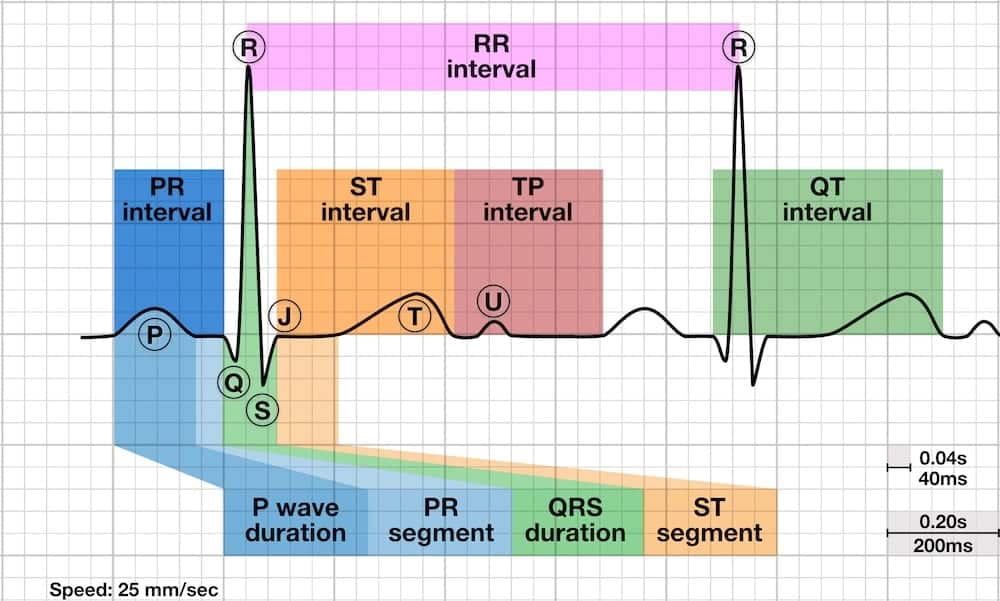
Vagal (parasympathetic) tone slows the resting sinus rate. Sympathetic activity, fever, pain, hypovolemia and hypoxia push it up. The rate also changes other measurements. As the rate climbs, the QT interval shortens, so use the corrected QT (see QT Interval). At fast rates, P waves can merge into the T wave before them (see P Wave). A short cycle after a long pause can conduct with a bundle-branch pattern (Ashman phenomenon, see Atrial Rhythms).
Normal Values & How to Measure
The normal sinus rate in adults is 60–100 bpm at rest. Pick the method by the rhythm. Box methods need a regular rhythm. Timed strip methods give an average, which is what you need for atrial fibrillation (AF) and other irregular rhythms.
| Method | Formula | Best For | Pitfall |
|---|---|---|---|
| 300 rule (large boxes) | 300 ÷ large boxes between R waves | Regular rhythms, quick estimate | Steps by value: 1 box = 300, 2 = 150, 3 = 100, 4 = 75, 5 = 60, 6 = 50 |
| 1500 rule (small boxes) | 1500 ÷ small boxes between R waves | Regular rhythms, precise rate | Many boxes to count; a miscount shifts the rate |
| 6-second strip | QRS complexes in 6 s (30 large boxes) × 10 | Irregular rhythms, slow rates | Gives an average; judge beats cut by the strip edges |
| 10-second strip | QRS complexes in 10 s (50 large boxes) × 6 | Very irregular rhythms | Longer count; still an average |
| R-R interval | 60 ÷ R-R interval in seconds | Single beats in a regular rhythm; digital ECG | One interval misleads in an irregular rhythm |
- Confirm calibration: standard paper speed is 25 mm/s (large box 0.2 s, small box 0.04 s). A different speed invalidates the box formulas.
- Pick the lead: use the longest rhythm strip, usually lead II, where R waves are tall and P waves are visible.
- Regular rhythm: count large boxes between two R waves and apply 300 ÷ boxes; check with the 1500 rule.
- Irregular rhythm: count QRS complexes in a 6-second window (30 large boxes) and multiply by 10.
- Count P waves separately: count P waves in the same window to compare atrial and ventricular rates in block or flutter.
- Report the method: for example, "Atrial fibrillation, average rate 85 bpm by 6-second count."
Sinus Bradycardia
Sinus bradycardia is a sinus rhythm slower than 60 bpm. Every QRS has a P wave in front of it. The PR interval and the QRS are normal.
ECG Criteria
- Rate: under 60 bpm, regular or varying with breathing.
- P waves: upright in lead II, one before each QRS (1:1 conduction).
- PR and QRS: PR 120–200 ms (0.12–0.20 s; 3–5 small boxes); QRS under 120 ms.
Causes
- Physiologic: sleep, endurance training and high vagal tone.
- Medications: beta-blockers, non-dihydropyridine calcium channel blockers, digoxin and clonidine.
- Metabolic and endocrine: hypothyroidism, hypothermia, hyperkalemia and hypoxia.
- Neurologic: raised intracranial pressure, which can produce a Cushing-type response.
- Cardiac: inferior myocardial infarction (MI), which can involve the AV node, and sinus node disease.
Clinical Significance
- Asymptomatic: usually no treatment. Find the cause and review medications.
- Symptomatic: hypotension, altered mental status, chest pain, heart failure or syncope. That needs treatment now (see Bedside Management & Escalation).
- Inferior MI: with a slow rate and inferior ST elevation, get a right-sided ECG. You are looking for right ventricular involvement.
Look-Alikes
- Sinus arrhythmia: the rate varies with breathing. That is a normal finding (see P Wave).
- AV block with a slow ventricular rate: the P waves and QRS complexes are not linked (see Conduction Blocks).
Sinus Pause, Sinus Arrest and Sick Sinus Syndrome
A sinus pause is a gap with no sinus P wave, and it runs longer than the usual P-P interval. Sinus arrest is the sinus node going quiet for a prolonged stretch. An escape rhythm may end the pause, or it may not.
ECG Criteria
- Pause: a P-P gap longer than the basic P-P interval, with no P wave inside it.
- Pause length: a pause that is an exact multiple of the basic P-P interval suggests sinus exit block; a pause that is not a multiple suggests sinus arrest.
- Escape: a narrow junctional escape or a wide ventricular escape may end the pause.
Causes
- Sinus node disease: age-related fibrosis is the most common cause.
- Medications: beta-blockers, calcium channel blockers, digoxin and antiarrhythmics.
- Vagal events: carotid sinus hypersensitivity, vasovagal reactions and carotid massage.
- Electrolytes and ischemia: hyperkalemia and inferior MI.
Sick Sinus Syndrome
Sick sinus syndrome is sinus node dysfunction that causes symptoms. It covers sinus bradycardia, pauses and sinus arrest. Often they alternate with a fast atrial rhythm: the tachy-brady pattern.
- Symptoms: dizziness, presyncope, syncope, fatigue or palpitations.
- Treatment: when symptoms match the bradycardia or pauses, permanent pacing is the standard treatment (2018 ACC/AHA/HRS bradycardia guideline). Review drugs that slow the sinus node.
Sinus Tachycardia
Sinus tachycardia is a regular sinus rhythm faster than 100 bpm, with a P wave in front of every QRS. It is a response, not a primary arrhythmia. Something is driving the heart: pain, fever or blood loss. Find the driver.
ECG Criteria
- Rate: above 100 bpm, typically rising and falling gradually.
- P waves: upright in lead II, one before each QRS; PR and QRS normal.
- Onset: gradual. An abrupt start or stop points to supraventricular tachycardia (SVT) instead.
Causes
- Volume loss: hypovolemia and hemorrhage.
- Infection and inflammation: sepsis and fever.
- Cardiopulmonary: heart failure, pulmonary embolism and hypoxia.
- Pain, anxiety and stimulants: including caffeine, sympathomimetic drugs and cocaine.
- Endocrine and drugs: hyperthyroidism, anemia, anticholinergic drugs and beta-agonists.
Clinical Significance
- Treat the cause: sinus tachycardia is a warning sign, and the rate usually falls when the driver is corrected.
- Do not slow it reflexively: slowing the rate can reduce cardiac output in a patient who is compensating.
Narrow- and Wide-Complex Tachycardia
Start tachycardia management with the QRS width, measured in the lead with the widest complex. Under 120 ms (under 0.12 s) is narrow. 120 ms or more is wide. Treat a regular wide-complex tachycardia as ventricular tachycardia (VT) until proven otherwise.
| Feature | Narrow QRS (under 120 ms) | Wide QRS (120 ms or more) |
|---|---|---|
| Common rhythms | Sinus tachycardia, AV nodal re-entrant or AV re-entrant tachycardia (AVNRT or AVRT), atrial flutter, AF, atrial tachycardia | VT, SVT with bundle branch block or rate-related aberrancy, pre-excited AF, torsades de pointes |
| Safe first assumption | Supraventricular origin, unless the rhythm or the patient says otherwise | VT, and pre-excited AF if the rhythm is irregular |
| Key clue | Regular rhythm with flutter waves or P waves in a fixed relation to the QRS | AV dissociation, capture or fusion beats, and a QRS unlike a bundle branch block |
- The 150 rule: regular, narrow and at about 150 bpm? It is atrial flutter with 2:1 block until proven otherwise.
- Bundle branch block (BBB): at fast rates, a BBB can make a wide QRS mimic VT. Compare with an old ECG, or with the same lead at a slower rate.
Rate in Irregular Rhythms
In an irregular rhythm, the rate is an average over time. Use a timed count of 6 or 10 seconds. One R-R interval is unreliable.
Atrial Fibrillation
- Pattern: irregularly irregular QRS complexes with no consistent P waves (see Atrial Rhythms).
- Rate: 6-second count × 10. In stable patients, a lenient rate-control target of under 110 bpm at rest is reasonable (2023 ACC/AHA/ACCP/HRS AF guideline).
- Rate classes: under 60 bpm (slow); 60–110 bpm (within the lenient resting target); over 110 bpm, or any rate with symptoms or hypotension (rapid ventricular response, RVR).
Atrial Flutter With Variable Block
- Pattern: atrial rate of about 250–350 bpm; the ventricular response depends on conduction (2:1 near 150 bpm, 4:1 near 75 bpm).
- Rate: a fixed block gives a regular ventricular rate. A variable block needs a timed count.
- Identify: sawtooth flutter waves, best seen in leads II, III, aVF and V1.
Ectopy and Bigeminy
- Rate: count every QRS for the total rate. Count sinus P waves separately for the underlying rate.
- Pulse deficit: premature beats that come too early to fill the ventricle may not give a palpable pulse. So the pulse can read lower than the ECG rate.
- Bigeminy: every other beat is premature. The palpated rate can fall well below the monitor rate.
Second-Degree AV Block (Mobitz I)
In Wenckebach block, the atrial rate is regular and faster than the ventricular rate. Count P waves and QRS complexes separately. The full pattern is on Conduction Blocks.
Reporting
- Average rate: "Atrial fibrillation, average rate 85 bpm."
- Rate range: "Atrial fibrillation, rate 70–110 bpm."
- Control: "Atrial fibrillation with RVR, rate 135 bpm."
Differential Diagnosis & Mimics
Rate alone rarely tells these rhythms apart. Look at onset, at the P waves, and at what vagal maneuvers or adenosine do.
| Feature | Sinus Tachycardia | Atrial Flutter (2:1) | Paroxysmal SVT (AVNRT or AVRT) |
|---|---|---|---|
| Onset and offset | Gradual | Often steady, may be paroxysmal | Abrupt start and stop |
| P waves | Upright in II, one before each QRS | Flutter waves near 300 per minute, sawtooth in II, III and aVF | Often hidden in or just after the QRS; retrograde P waves may be inverted in II, III and aVF |
| Vagal maneuver or adenosine | Slows gradually; no termination | Transient slowing unmasks flutter waves | Terminates abruptly in AVNRT and AVRT |
Wide-Complex Mimics
- Ventricular tachycardia vs SVT with aberrancy: look for AV dissociation, capture or fusion beats. Precordial morphology that does not fit a bundle branch block also counts (criteria described by Brugada).
- Pre-excited AF: irregular, wide and very fast, with QRS shapes that keep changing (see the warning in Bedside Management & Escalation).
Artifact & Lead Misplacement
- Limb lead reversal: an arm swap inverts lead I. It can mimic dextrocardia or an abnormal axis (see Cardiac Axis).
- Muscle tremor and shivering: can look like AF or flutter. Check the pulse and the SpO₂ waveform before you treat the strip.
- Transport vibration: can mimic a fast or wide rhythm. Recheck the strip when the vehicle is stationary.
- Electrode drift: baseline wander or poor contact can hide P waves. It can also make R-R intervals unmeasurable.
Normal Variants
- Sinus arrhythmia: the rate rises with inspiration and falls with expiration. It is common in young adults and benign.
- Athletic bradycardia: resting sinus bradycardia is a recognized adaptation in trained athletes. Symptoms and context decide whether it needs a work-up (International Criteria for ECG interpretation in athletes).
- Sleep: the sinus rate falls during sleep.
Bedside Management & Escalation
Rate decisions follow perfusion. Not the number alone. Use the stability table to pick the pathway, and the therapy table for doses. Pediatric Advanced Life Support (PALS) doses sit in the pediatric column.
| Situation | BLS (EMT) | ALS (AEMT / Paramedic / RN) | Critical Care / Transport |
|---|---|---|---|
| Unstable bradycardia (under 50 bpm with poor perfusion) | Oxygen if SpO₂ under 94%; monitor; call ALS; apply pads | Atropine; if ineffective, transcutaneous pacing and/or epinephrine or dopamine infusion, per the AHA 2025 adult bradycardia algorithm | Pacing ready early; confirm capture by pulse; treat hypoxia and toxins |
| Stable bradycardia | Monitor; 12-lead where allowed; transmit | Observe and treat the cause; atropine for symptomatic bradycardia (1 mg IV every 3–5 min, max 3 mg; see table) | Serial 12-lead; notify; pacing on standby |
| Unstable tachycardia with a pulse | Oxygen; monitor; call ALS; apply pads | Synchronized cardioversion (see table); sedate whenever feasible (AHA 2025 adult tachyarrhythmia algorithm, box 6), without delaying the shock (AHA 2025 pediatric tachyarrhythmia algorithm) | Synchronized cardioversion; reassess after each shock |
| Stable narrow regular tachycardia | Monitor; vagal maneuver per protocol | Vagal maneuver, then adenosine | Adenosine with monitoring; 12-lead; cardioversion ready |
| Stable wide-complex tachycardia | Treat as VT; call ALS | Amiodarone or procainamide (see table); cardiovert if unstable | Synchronized cardioversion if unstable; reassess after each shock |
| Pulseless (any rhythm) | CPR and AED | Adult or pediatric arrest algorithm | Arrest pathway; termination rules per protocol |
| Therapy | Adult | Pediatric | Key Notes |
|---|---|---|---|
| Atropine | 1 mg IV/IO every 3–5 min (max 3 mg) | 0.02 mg/kg IV/IO (min 0.1 mg, max 0.5 mg per dose); may repeat once | For symptomatic bradycardia. Label antivagal dose is 0.5–1 mg (Dosage and Administration); the label 1 mg dose is for bradyasystolic arrest, with 3 mg maximum total. Use cautiously in acute MI or ischemia, where the faster rate can worsen ischemia. May be ineffective in infranodal block and in a transplanted heart; do not delay pacing. |
| Transcutaneous pacing (pacing page) | Rate 60–80/min; increase output until capture, about 10% above threshold | Specialist use; pediatric pads or attenuator per device | Confirm capture by pulse or SpO₂ waveform, not by the pacing spike alone. Sedation may be needed. |
| Epinephrine High-alert | Bradycardia: 2–10 mcg/min IV infusion, titrated | 0.01 mg/kg IV/IO every 3–5 min (max 1 mg per dose); infusion 0.1–1 mcg/kg/min for shock | Check concentration (1 mg/mL vs 0.1 mg/mL) and units (mcg/min vs mcg/kg/min) before each dose or infusion. |
| Dopamine High-alert | Bradycardia: 5–20 mcg/kg/min IV infusion, titrated (max 20 mcg/kg/min) | 2–20 mcg/kg/min IV infusion, titrated to heart rate and blood pressure | AHA 2025 adult bradycardia recommendation (COR 2b, LOE C-LD): if atropine fails, epinephrine, dopamine or transcutaneous pacing may be effective; the choice depends on clinician experience and resources. |
| Adenosine | 6 mg rapid IV push with flush; second dose 12 mg rapid IV push if required, per the AHA 2025 adult tachyarrhythmia algorithm. Max 12 mg per dose. | 0.1 mg/kg rapid IV/IO (max 6 mg), then 0.2 mg/kg (max 12 mg) | Narrow regular SVT only. Warn of flushing and brief asystole. Central line or post-heart transplant: effects can be profound. Per the AHA 2025 adult narrow-complex tachycardia text, 1 mg IV may be adequate. Smaller doses may work with dipyridamole; carbamazepine can increase heart block. Caffeine and theophylline antagonize it, so larger doses may be needed. Avoid in bronchospasm or asthma; use caution in COPD. Effects are prolonged in a transplanted heart. |
| Synchronized cardioversion | Narrow-complex tachycardia: 100 J. Atrial fibrillation: at least 200 J biphasic as the first setting; atrial flutter: 200 J (per the AHA 2025 adult recommendations, AF COR 2a, LOE B-R). Monomorphic VT with a pulse: 100 J. Increase stepwise per device and protocol. | 0.5–1 J/kg, then 2 J/kg if the first shock fails | Confirm the sync marker on every R wave. Pulseless VT or VF is defibrillation, not cardioversion. |
| Amiodarone High-alert | Pulseless VT or VF: 300 mg IV/IO first dose, then 150 mg second dose. Stable VT: 150 mg IV over 10 min, then 1 mg/min for the first 6 hr (AHA 2025 ACLS tachyarrhythmia algorithm) | Pulseless VT or VF: 5 mg/kg rapid IV/IO (max 300 mg first dose); may repeat up to 3 doses, max 150 mg for later doses. Perfusing VT: specialist use. | Watch for hypotension and bradycardia during the load. Not for torsades de pointes; give magnesium first. |
| Procainamide High-alert | Stable wide-complex tachycardia: 20–50 mg/min IV to an endpoint (max 17 mg/kg) | Specialist use; no PALS dose | Stop for hypotension, QRS widening over 50%, or the maximum. Avoid with prolonged QT or heart failure. |
| Diltiazem | AF with RVR, hemodynamically stable: 0.25 mg/kg IV over 2 min (about 20 mg); if response is inadequate after 15 min, 0.35 mg/kg (about 25 mg). Then infusion at 10 mg/hr, titrated 5–15 mg/hr. | Specialist use only; not established in children | Not for pre-excited AF or wide-complex tachycardia. Avoid in decompensated heart failure with reduced EF (LVEF under 40%), hypotension or shock; unstable AF needs synchronized cardioversion. Do not give with IV beta-blockers within a few hours. Watch blood pressure. |
Where local protocol sets a different dose, this differs from the AHA algorithm.
Pediatric & Special Populations
Pediatrics
| Age | Median Heart Rate (bpm) | Notes |
|---|---|---|
| Birth | 127 | Median from a systematic review of pediatric ranges |
| About 1 month | 145 | Peak of the median curve |
| 2 years | 113 | Falls through childhood; use the age table at Pediatric Vital Signs |
- Tachycardia in a child: sinus tachycardia is typically under 220 bpm in infants and under 180 bpm in children. Expect a P wave before each QRS and variable R-R intervals. A narrow-QRS rate typically at or above these values, with little beat-to-beat variability, suggests SVT (per the AHA 2025 pediatric tachyarrhythmia algorithm). For the age-specific normal range, see Pediatric Vital Signs.
- Bradycardia in a child: it usually reflects hypoxia or poor perfusion. A heart rate under 60 bpm with poor perfusion, despite oxygenation and ventilation, needs CPR, IV/IO access and epinephrine. Atropine is for increased vagal tone or primary AV block (per the AHA 2025 pediatric bradycardia algorithm).
- Pediatric drug doses: the weight-based atropine, epinephrine and adenosine doses are in the Bedside Management table. Check the weight-based math on Weight-Based Dosing.
- Measurement: young children move and cry. Get the strip from a calm child, and do not read a crying rate as baseline.
Other Populations
- Pregnancy: resting sinus tachycardia is common. Blood volume and cardiac output rise, so judge a new tachycardia by context, not by rate alone. Manage unstable arrhythmias as in non-pregnant adults. Cardioversion is safe. After about 20 weeks, displace the uterus to the left (left lateral tilt or manual displacement) during resuscitation or transport. A new tachycardia or bradycardia can signal hemorrhage, pulmonary embolism or peripartum cardiomyopathy.
- Older adults: sinus node and AV conduction disease are more common. Pauses and bradycardia may be the first sign of ischemia or drug toxicity.
- Athletes: resting sinus bradycardia is common. It is usually benign (see Normal Variants).
- Heart transplant: the denervated heart does not respond to atropine. Adenosine effects can be prolonged.
- Pacemaker or ICD: know the programmed lower rate. A rate below it points to failure to capture or failure to sense (see Ventricular Rhythms).
Monitoring & Transport Considerations
| Issue | What to Do |
|---|---|
| Rhythm lead | Use lead II for the rhythm. Use V1 or the monitor lead that shows the QRS shape for bundle branch block and wide-complex analysis. |
| Artifact | Shivering, movement and transport vibration can mimic tachycardia or VT. Check the pulse and SpO₂ waveform before treating. |
| Pads | Place anterior–posterior pads before moving an unstable slow or fast patient. |
| Pacing | Confirm capture by pulse or SpO₂ waveform. The pacing spike shows output, not mechanical capture. |
| Reassessment | Repeat the rate, rhythm and blood pressure after each intervention and after each move. |
| Pacemaker | Compare the rate with the programmed lower limit. A rate below it suggests failure to capture or inhibition (see Ventricular Rhythms). |
| Medication alarms | Set heart rate alarms for infusions. Watch for hypotension and bradycardia with amiodarone, diltiazem and beta-blockers. |
| Handoff | Give the rate, the calculation method, the rhythm, the time of each intervention and the response. |
Step-by-Step Approach & Pitfalls
- Check responsiveness, pulse, blood pressure and SpO₂ before reading the monitor number.
- Obtain a 6-second rhythm strip in lead II and confirm the paper speed.
- Classify the rate: under 60 bpm (slow), 60–100 bpm (normal), or over 100 bpm (fast).
- Decide whether the rhythm is regular or irregular, then calculate with the method that fits.
- Count P waves against QRS complexes to find the atrial rate and any block.
- For a fast rhythm, measure the QRS width in the widest lead: narrow is under 120 ms; wide is 120 ms or more.
- Decide stability from mental status, blood pressure, chest pain, heart failure and shock, then follow the stability table.
- Repeat the rhythm strip and perfusion check after each intervention, and document rate, rhythm, method and response.
Common Pitfalls
- Box rules on irregular rhythms: the 300 and 1500 rules have no place in AF.
- Calculating from one R-R interval: one interval misstates the rate in AF and in ectopy.
- Treating the number, not the patient: 45 with normal mental status and blood pressure is not the same as 45 with hypotension.
- Missing flutter at 150: a regular narrow tachycardia near 150 bpm can turn out to be 2:1 flutter.
- Missing hidden P waves: at fast rates, P waves can hide inside the T wave or the QRS.
- Missing a drug cause: beta-blockers, calcium channel blockers and digoxin account for many slow rates.
- Trusting the pulse: a palpated rate can run lower than the ECG rate because of a pulse deficit.
Clinical Pearls
References
- Wigginton JG, et al. (2025). Part 9: Adult advanced life support: 2025 American Heart Association Guidelines for Cardiopulmonary Resuscitation and Emergency Cardiovascular Care. Circulation. 2025;152(16 suppl 2). https://doi.org/10.1161/CIR.0000000000001376
- Lasa JJ, et al. (2025). Part 8: Pediatric advanced life support: 2025 American Heart Association and American Academy of Pediatrics Guidelines for Cardiopulmonary Resuscitation and Emergency Cardiovascular Care. Circulation. 2025;152(16 suppl 2). https://doi.org/10.1161/CIR.0000000000001368
- Kusumoto FM, et al. (2019). 2018 ACC/AHA/HRS guideline on the evaluation and management of patients with bradycardia and cardiac conduction delay. Circulation. 2019;140(8). https://doi.org/10.1161/CIR.0000000000000628
- Joglar JA, et al. (2024). 2023 ACC/AHA/ACCP/HRS guideline for the diagnosis and management of atrial fibrillation. Circulation. 2024;149(1):e1–e156. https://doi.org/10.1161/CIR.0000000000001193
- Kligfield P, et al. (2007). Recommendations for the standardization and interpretation of the electrocardiogram (landmark standardization statement). Circulation. 2007;115:1306–1324. https://doi.org/10.1161/CIRCULATIONAHA.106.180200
- Fleming S, et al. (2011). Normal ranges of heart rate and respiratory rate in children from birth to 18 years of age: a systematic review of observational studies (landmark systematic review). Lancet. 2011;377(9770):1011–1018. https://doi.org/10.1016/S0140-6736(10)62226-X
- Drezner JA, et al. (2017). International criteria for electrocardiographic interpretation in athletes: consensus statement. British Journal of Sports Medicine. 2017. https://doi.org/10.1136/bjsports-2016-097331
- Hospira, Inc. (2025). Atropine sulfate injection, for intravenous use [prescribing information; version 30, revised Nov 17, 2025]. DailyMed, National Library of Medicine. https://dailymed.nlm.nih.gov/dailymed/drugInfo.cfm?setid=ad8916e7-206e-409e-2582-30d072845dd4
- American Heart Association (2025). 2025 American Heart Association Guidelines for Cardiopulmonary Resuscitation and Emergency Cardiovascular Care, Part 9: Adult Advanced Life Support. Initial bradycardia management (COR 2b, LOE C-LD); synchronized cardioversion of atrial fibrillation with at least 200 J biphasic (COR 2a, LOE B-R); adenosine in central-line and post-transplant patients. https://cpr.heart.org/en/resuscitation-science/cpr-and-ecc-guidelines/adult-advanced-life-support
- American Heart Association (2025). 2025 American Heart Association Guidelines for CPR and ECC: Algorithms, including adult bradycardia, adult tachyarrhythmia, electrical cardioversion, pediatric bradycardia and pediatric tachyarrhythmia with a pulse, and pediatric cardiac arrest doses. https://cpr.heart.org/en/resuscitation-science/cpr-and-ecc-guidelines/algorithms
Medical Disclaimer
- For Educational Purposes Only: This content is intended for educational reference and should not be used for clinical decision-making.
- Not a Substitute for Professional Judgment: Always consult your local protocols, institutional guidelines, and supervising physicians.
- Verify Before Acting: Users are responsible for verifying information through authoritative sources before any clinical application.