

At a Glance
- Pattern
- Sudden-death ECG patterns hidden in a syncope workup: Brugada, long QT, pre-excitation, ARVC and HCM. A normal-looking ECG does not always rule them out.
- Normal (Adult)
- QTc <450 ms (men), <460 ms (women); PR 120–200 ms; no coved ST elevation in V1–V2; QRS ≤110 ms; no epsilon wave.
- Confirm It
- QTc in lead II or V5–V6 over several beats; PR and delta wave in II; V1–V2 one or two spaces higher if standard leads look saddleback.
- Abnormal → Think
- Coved V1–V2 ST elevation: Brugada, fever or drug unmasking. Short PR with wide QRS: pre-excitation. T inversion in V1–V3 with epsilon waves: ARVC. Long QTc: QT drugs or low K⁺, Mg²⁺, Ca²⁺.
- Red Flags
- Type 1 Brugada with syncope; QTc ≥500 ms or a 60 ms rise; irregular wide-complex tachycardia with pre-excitation; any abnormal ECG with syncope.
- Bedside Action
- Stop QT-prolonging and sodium-channel-blocking drugs; magnesium 2 g IV for torsades; unsynchronized shock if pulseless; no AV-nodal blockers or IV amiodarone in pre-excited AF; cardiology or EP for every high-risk pattern.
Physiology & What It Represents
Most syncope is benign. A small group of ECG patterns points to an arrhythmic cause, and that cause can end in sudden cardiac death. The 2017 ACC/AHA/HRS and 2018 ESC syncope guidelines both put the 12-lead ECG in the initial evaluation. A 12-lead is at the bedside within minutes.
All five patterns on this page have one thing in common. They show on a standard 12-lead before any arrhythmia is recorded. Each one has its own mechanism.
- Brugada: An action-potential notch on the epicardium of the right ventricle, not the endocardium, sets up a voltage gradient. That gradient shows as a J wave or raised J point in V1–V2. Sodium-channel blockade makes the phase-1 notch more pronounced.
- Pre-excitation: an accessory pathway goes around the AV node, so the ventricles get the signal early. That shortens the PR and slurs the QRS upstroke (the delta wave).
- Long QT: prolonged repolarization sets the stage for torsades de pointes, a polymorphic VT.
- ARVC and HCM: structural disease leaves its mark. In ARVC, that is repolarization change and delayed activation in the right precordial leads. In HCM, that is left ventricular hypertrophy voltage.
Other high-risk ECG features in syncope
The 2017 ACC/AHA/HRS and 2018 ESC guidelines also read these ECG findings as pointing to an arrhythmic cause. This page does not go deep on them. Use the linked E3 pages:
- High-grade AV block (Mobitz II or third-degree) and severe sinus bradycardia or pauses over 3 s: see Conduction Blocks and Sinus Rhythms.
- Bifascicular or other conduction disease: see Conduction Blocks.
- Ischemia, pathologic Q waves or acute MI: see Q Wave and STEMI Recognition.
- Nonsustained or sustained VT: see Ventricular Rhythms.
"Abnormal ECG" on this page means one of these features or one of the patterns below. It does not mean any minor variant.
Normal Values & How to Measure
| Parameter | Adult Normal | Pediatric Note | Where / How to Measure |
|---|---|---|---|
| QTc | <450 ms (men), <460 ms (women) | Age-dependent; see the QT Interval page | Lead II or V5–V6, averaged over several beats; Fridericia at HR <60 or >90 bpm |
| PR interval | 120–200 ms (3–5 small boxes) | Shorter in infants | Lead II; a PR under 120 ms with a delta wave suggests pre-excitation |
| QRS duration | ≤110 ms (111–119 ms borderline) | See the QRS Complex page | Widest lead; ≥120 ms is wide |
| J point and ST, V1–V2 | No coved ST elevation ≥2 mm | See Pediatric & Special Populations | Standard position, then one and two intercostal spaces higher |
| T wave, V1–V3 | Upright | Inverted V1–V3 can be normal through adolescence | Inversion in V1–V3 is an ARVC criterion when complete RBBB is absent |
| Epsilon wave | None | — | Small, reproducible signal between the end of the QRS and the T wave in V1–V3 (major ARVC criterion) |
- Standardize the tracing: 25 mm/s and 10 mm/mV. Record durations in ms first, then seconds and small boxes.
- Measure QTc: QRS onset to T-wave end in lead II or V5–V6, averaged over several beats, corrected with Fridericia when the rate is outside 60–90 bpm.
- Measure the J point: record ST elevation at the J point in millimeters for V1 and V2.
- Repeat V1–V2 higher: if standard V1–V2 show a saddleback pattern, record one and two intercostal spaces higher, then compare.
- Look for a delta wave: check the PR interval and the QRS upstroke in lead II and the precordial leads.
- Scan V1–V3 for epsilon waves: a reproducible notch after the QRS and before the T wave.
Brugada Syndrome (Type 1 and Type 2)
Brugada syndrome is an inherited channelopathy. It sets up structurally normal hearts for polymorphic ventricular tachycardia, ventricular fibrillation and sudden cardiac death. The ECG shows a right precordial ST pattern. In some patients it is there all the time. In others, drugs or fever bring it out.
ECG Criteria
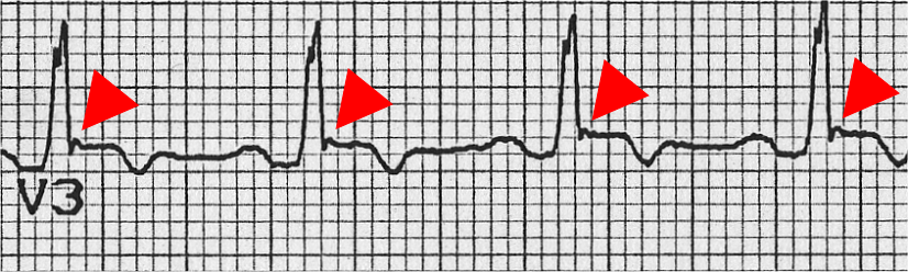
- Type 1 (coved): ST elevation of 2 mm or more followed by a negative T wave, in at least one of V1–V2 at the standard position or one or two intercostal spaces higher. This is the diagnostic pattern.
- Type 2 (saddleback): J-point elevation of 2 mm or more with ST elevation of 1 mm or more. Suggestive, not diagnostic on its own; a sodium-channel blocker challenge is used to look for conversion to type 1.
- Diagnosis: a type 1 pattern in at least one of V1–V2 (standard or high position), spontaneous or after a sodium-channel blocker challenge, establishes Brugada syndrome. Symptoms determine risk and management.
Causes
- Fever: It can unmask a type 1 pattern. The pattern usually disappears once the patient is afebrile, but arrhythmic events have been reported during fever.
- Sodium-channel blockers: Flecainide and procainamide can unmask a type 1 pattern. Use them only as a monitored electrophysiology challenge. Before you give any new drug, check the Brugada drug list at brugadadrugs.org.
- Alcohol and cannabis: they sit on the same list as fever and sodium-channel blockers, as triggers to avoid.
Clinical Significance
- Risk: A type 1 pattern with syncope is the high-risk setting. You do not need structural heart disease to make the diagnosis.
- Storm: For Brugada or VF storm, isoproterenol is a specialist option, used after magnesium and defibrillation (see Bedside Management). It is not a first-line Brugada drug.
- Next step: Get urgent cardiology or electrophysiology review. That team makes the implantable defibrillator decision after a shared-decision discussion.
Look-Alikes
- Right bundle branch block, and high-placed V1–V2 leads (see Differential Diagnosis & Mimics).
Long QT Syndrome and Torsades Risk
Long QT, whether acquired or congenital, delays repolarization. The dangerous end point is torsades de pointes. It is a polymorphic ventricular tachycardia with a long QT, and it presents as syncope or sudden arrest.
ECG Criteria
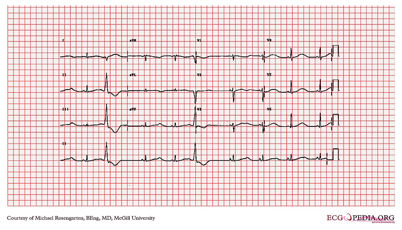
- Prolonged: QTc ≥450 ms in men or ≥460 ms in women.
- High torsades risk: QTc ≥500 ms, or a rise of 60 ms or more from the baseline value.
- Syncope with a long QT is a red flag on its own, even when the QTc is only moderately prolonged.
Causes
- Drugs: many QT-prolonging drugs, including antiarrhythmics, antipsychotics, some antiemetics, methadone and some antibiotics (see the QT Interval page).
- Electrolytes: low K⁺, Mg²⁺ or Ca²⁺.
- Rhythm: bradycardia or pauses, hypothermia, ischemia and raised intracranial pressure.
- Congenital long QT: think about it when no acquired cause turns up.
Clinical Significance
- Torsades: Magnesium given IV may suppress recurrent torsades regardless of serum Mg²⁺ (AHA: may be considered). For unstable torsades, with or without a pulse, you defibrillate unsynchronized.
- Congenital triggers: LQT1, exertion or swimming; LQT2, sudden auditory stimuli or startle; LQT3, rest or sleep.
- Pause-dependent torsades: overdrive pacing or isoproterenol, per specialist direction.
Look-Alikes
- A wide QRS, from bundle branch block or ventricular pacing, makes the QT look longer than it is. Measure the QT in a lead with a clear T-wave end, or use the method on the QT Interval page.
Pre-Excitation (WPW) and Pre-Excited AF
Pre-excitation is the ventricles getting activated early through an accessory pathway that skips the AV node. In atrial fibrillation, that pathway can carry impulses very fast, and the ventricles can go into fibrillation.
ECG Criteria
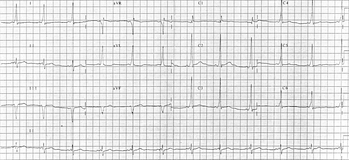
- Delta wave: slurred QRS upstroke with a short PR interval (under 120 ms) in sinus rhythm.
- Pre-excited AF: an irregular, very fast, wide-complex tachycardia with broad and changing QRS complexes.
- High risk: a shortest pre-excited R–R interval under 250 ms marks a pathway that can conduct fast.
Causes
- Congenital accessory pathway: This is the usual cause of pre-excitation.
- Intermittent pre-excitation: The delta wave can come and go. One normal ECG does not exclude a pathway.
Clinical Significance
- Mimic: Pre-excited AF can pass for SVT or VT. Give it AV-nodal blockers or IV amiodarone and it can collapse the patient or go to VF.
- Definitive care: Electrophysiology arranges catheter ablation of the accessory pathway.
Look-Alikes
- Short PR without delta wave (see the PR Interval page) and wide-complex tachycardia of other causes (see Differential).
Arrhythmogenic Right Ventricular Cardiomyopathy (ARVC)
Arrhythmogenic right ventricular cardiomyopathy is an inherited disease of the right ventricle. It drives ventricular arrhythmias and sudden death. The ECG is one input among several. Imaging, histology, genetics and family history count too.
ECG Criteria
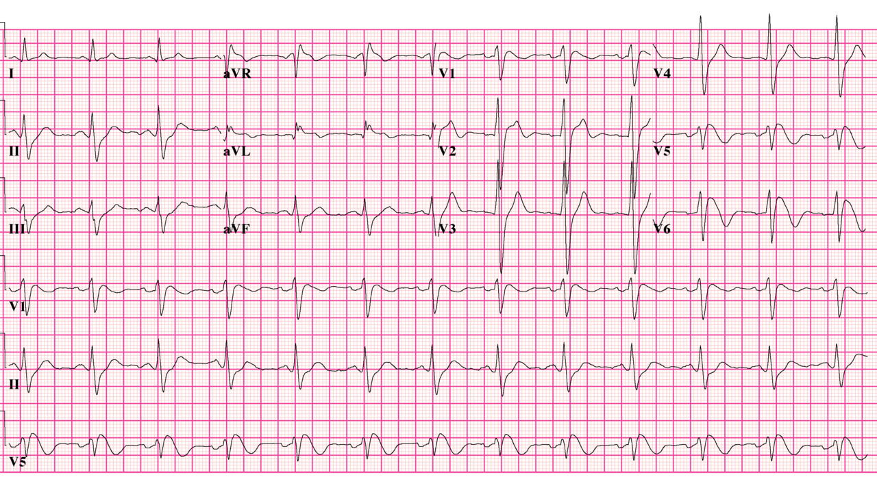
- T-wave inversion in V1–V3 (adults and children over 14 years): a major repolarization criterion in the 2010 Task Force Criteria when complete RBBB is absent. With complete RBBB, inversion in V1–V4 is a minor criterion. Without RBBB, inversion in V1–V2 or V4–V6 is also a minor criterion.
- Epsilon waves (major criterion): reproducible low-amplitude signals between the end of the QRS and the start of the T wave in V1–V3.
- Terminal activation duration of the QRS of 55 ms or more in V1–V3 (minor criterion), when complete RBBB is absent.
- Ventricular tachycardia with LBBB morphology and a superior axis: a recognized ARVC pattern.
Causes
- It is an inherited right-ventricular disease. ECG findings are one part of a multi-factor diagnosis.
Clinical Significance
- Accuracy: When you separate ARVC from idiopathic outflow-tract ventricular arrhythmia, pooled T-wave inversion in V1–V3 showed a sensitivity of 0.55 and a specificity of 0.88. Pooled epsilon waves showed a sensitivity of 0.12 and a specificity of 0.98.
- Meaning: An epsilon wave is specific but rare. A negative scan does not exclude ARVC. Refer for cardiology review when the pattern shows up with syncope.
Look-Alikes
- Idiopathic outflow-tract ventricular arrhythmia and complete RBBB, which changes how the T-wave criterion is scored.
Hypertrophic Cardiomyopathy (HCM)
Hypertrophic cardiomyopathy is a myocardial disease. The left ventricle is thick, and loading conditions do not explain the wall thickness. In adults, the diagnosis uses a wall thickness of 15 mm or more, or 13–14 mm with a family history, a genetic finding or ECG abnormalities.
ECG Criteria
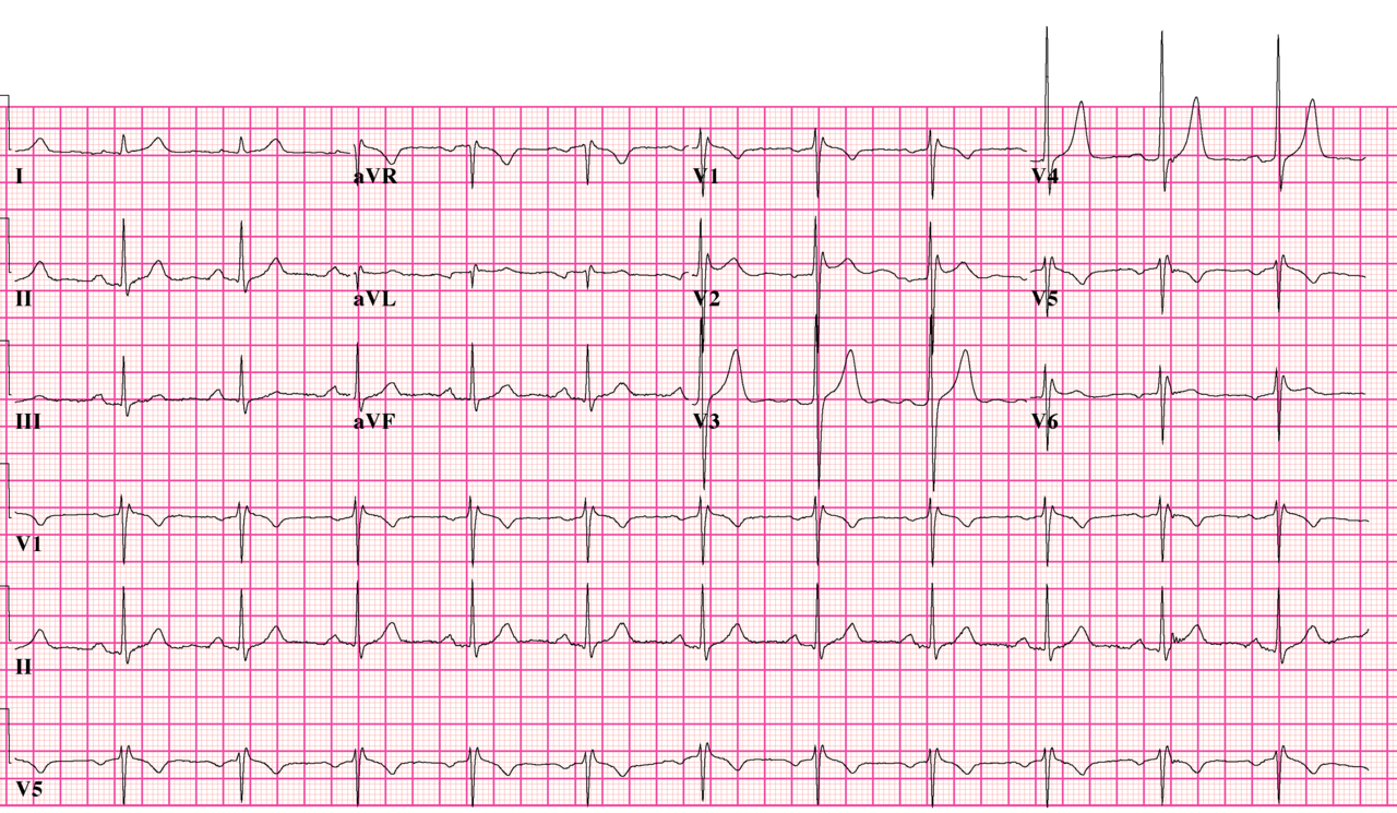
- Left ventricular hypertrophy: increased QRS voltage. It is usually present but seldom the only finding.
- Deep T-wave inversion in the lateral leads is typical of apical HCM.
- Q waves: can reflect fibrosis or segmental hypertrophy.
Causes
- Sarcomeric (genetic) HCM: This is the genetic form of the disease.
- Phenocopies: Cardiac amyloidosis and Fabry disease can look like HCM on the ECG and on imaging.
Clinical Significance
- Sudden death risk: The risk runs up to 0.5–2% per year in people with HCM. Non-sustained VT is an independent risk factor. Unexplained syncope is a variable in sudden-death risk models.
- Presentation: exertional dyspnea, chest pain, syncope or palpitations, sometimes with a murmur from left ventricular outflow tract obstruction.
- Next step: With suspected obstructive HCM and hypotension, avoid vasodilators (such as nitrates), inotropes and hypovolemia. Give volume and a pure vasoconstrictor such as phenylephrine, per medical direction. Notify cardiology early (2024 AHA/ACC HCM guideline, reference 5).
Differential Diagnosis & Mimics
Brugada, pre-excitation, long QT and the structural patterns can pass for one another. They can also pass for other causes of wide-complex or ST-changing ECGs. Compare them in this order.
| Feature | Brugada Type 1 | Brugada Type 2 | Right Bundle Branch Block |
|---|---|---|---|
| ST pattern in V1–V2 | Coved, ≥2 mm J-point ST elevation, then a negative T | Saddleback, J point ≥2 mm with ST ≥1 mm | QRS ≥120 ms (see QRS Complex) |
| Diagnostic alone? | Yes, with or without symptoms (symptoms set risk) | No; a challenge is used to look for conversion to type 1 | No; a conduction block, not a channelopathy |
| What changes it | Fever, sodium-channel blockers, alcohol, cannabis | Can convert to type 1 with a sodium-channel blocker | Persistent conduction pattern; compare with prior ECG |
Artifact & Lead Misplacement
- High V1–V2 placement can produce a coved pattern at higher intercostal spaces, one that is absent at the standard position. Write down where the leads sat.
- Limb-lead reversal changes the axis and can pass for a pathologic axis (see Cardiac Axis).
- Fever and drugs change the ST shape with no structural cause behind it. If the clinical picture allows, repeat the tracing afebrile and off the drug.
Normal Variants
- Small right precordial variations in healthy people can pass for a type 2 pattern. Judge them by the criteria above, not by how they look.
Bedside Management & Escalation
The stability table covers the three situations on this page. Doses appear only in the table below. Every row depends on protocol.
| Situation | BLS (EMT) | ALS (AEMT / Paramedic / RN) | Critical Care / Transport |
|---|---|---|---|
| Unstable: pulseless VF, pVT or torsades | CPR; AED shock. Pulseless torsades needs an unsynchronized shock | Magnesium 2 g IV for torsades, with or without a pulse; pulseless VF or pVT: shock and ALS protocol. If torsades or long QT is suspected, give magnesium and defibrillate; avoid amiodarone and procainamide. Keep K⁺ per protocol (often above 4 mmol/L) | Overdrive pacing or isoproterenol for pause-dependent torsades (specialist) |
| Unstable: pre-excited AF | 12-lead, oxygen if SpO₂ <94%, early ALS | Synchronized cardioversion per the Cardioversion page; no adenosine, diltiazem, verapamil, beta-blockers, digoxin or IV amiodarone | EP consult; continuous monitoring during transport |
| Stable, high-risk pattern | 12-lead, transmit if permitted, no drugs | IV access, monitor, notify; avoid QT-prolonging and sodium-channel-blocking drugs | Telemetry, serial ECGs, cardiology handoff; treat fever per protocol |
| Therapy | Adult | Pediatric | Key Notes |
|---|---|---|---|
| Magnesium sulfate (torsades) | 2 g IV/IO over about 15 min for torsades, with or without a pulse (after the unsynchronized shock if pulseless), repeat 2 g if episodes persist; no fixed max | 25–50 mg/kg IV/IO, max 2 g per dose | High-alert Give regardless of serum Mg²⁺ (may suppress recurrent torsades). On repeat doses monitor patellar reflexes, respiratory rate and BP; reduce in renal failure. Stop QT drugs and correct K⁺. Magnesium Sulfate |
| Isoproterenol (specialist-directed, off-label: pause-dependent torsades or VF storm) | 0.5–10 mcg/min IV infusion, titrated to heart rate, reduce if heart rate exceeds goal or 130/min | Not established in children | High-alert After magnesium and defibrillation; specialist use only. Do not give for congenital long QT, angina or ACS, tachyarrhythmia, or digoxin toxicity. Caution in hypertrophic cardiomyopathy with outflow obstruction (see HCM). Isoproterenol |
| Procainamide (stable pre-excited AF) | 20–50 mg/min IV to an endpoint, then 1–4 mg/min, max 17 mg/kg; label max 1 g load | Specialist use; see the Procainamide page for weight-based dosing | High-alert Off-label for pre-excited AF (AHA 2025 COR 2b). Record baseline QRS and BP; continuous ECG and BP every few minutes. Stop for hypotension (fall of 15 mmHg or more), QRS widening over 50%, arrhythmia suppressed, or max dose. Do not give with prolonged QT or torsades, heart failure, complete or high-grade AV block, SLE, or known or suspected Brugada. Not after amiodarone or ibutilide. Procainamide |
Doses vary by protocol; follow local protocols and medical direction.
- Brugada, suspected: stay away from sodium-channel blockers (flecainide, procainamide). Treat fever promptly per protocol.
- Syncope with a high-risk pattern: transfer to a monitored setting. Cardiology and electrophysiology decide on ICD, ablation and pacing.
Follow local protocols and online medical direction.
Pediatric & Special Populations
Pediatrics
- Red flags in children: syncope during exertion or while supine, no prodrome, palpitations or chest pain, an abnormal ECG or vital signs, and a family history of sudden cardiac death under 40 years all count as red flags. They are the high-risk features in the pathway.
- Disposition: every child with syncope gets a 12-lead ECG as part of the initial evaluation. High-risk children go to telemetry and echocardiography.
- Age-based QTc: read the QTc against age-appropriate thresholds on the QT Interval page.
- Doses: magnesium 25–50 mg/kg (max 2 g per dose). Isoproterenol is not established in children. See Weight-Based Dosing.
Other Populations
- Athletes: syncope during exercise counts as a high-risk feature, even when the patient is young and fit.
- Febrile patients: fever can unmask a Brugada pattern. Repeat the ECG once the fever breaks.
- Drug and substance users: find out about sodium-channel blockers, QT-prolonging drugs, alcohol and cannabis.
- Known cardiomyopathy or channelopathy: device settings belong to the treating cardiology team. A shock or syncope in a patient with an ICD calls for urgent device interrogation.
- Pregnancy: QT prolongation and arrhythmia risk shift around pregnancy and the postpartum period. Check the QTc, and bring in cardiology and obstetrics. For drug and cardioversion safety in pregnancy, see the Procainamide page.
Monitoring & Transport Considerations
| Issue | What to Do |
|---|---|
| Monitoring lead | Lead II for P waves and delta waves; V1–V2 at the standard and higher positions for Brugada; the tallest T-wave lead for QT trending |
| Serial ECGs | Repeat the 12-lead with fever, a new drug, or a change in symptoms. Brugada patterns can alternate between type 1 and type 2 |
| Telemetry | A high-risk pattern with syncope needs telemetry and echocardiography, usually admission |
| QT-prolonging drugs | Check the QTc before ondansetron or droperidol. Keep K⁺ and Mg²⁺ replaced |
| Sodium-channel blockers | Avoid flecainide and procainamide in known or suspected Brugada. A monitored electrophysiology challenge is the only setting for them |
| Pre-excited AF in transport | No AV-nodal blockers and no IV amiodarone. Cardioversion equipment ready; see the Cardioversion page |
Step-by-Step Approach & Pitfalls
- Take the syncope history: exertion, supine onset, prodrome, palpitations, chest pain and family history of sudden death under 40 years. For a long QT, ask about exertion or swimming, startle or sudden noise, and syncope at rest or sleep.
- Obtain a 12-lead ECG and compare it with any prior tracing.
- Measure the QTc in lead II or V5–V6, and check K⁺ and Mg²⁺.
- Look for a delta wave with a PR under 120 ms, and for an irregular wide-complex rhythm.
- Inspect V1–V3: coved or saddleback ST elevation, and epsilon waves. Record one and two spaces higher if the standard position looks saddleback.
- Ask about triggers: fever, alcohol, cannabis, sodium-channel blockers and QT-prolonging drugs.
- Hand off with the tracing: label the high-risk feature on the handoff, and do not discharge on a normal-looking ECG until triggers are reviewed.
Common Pitfalls
- Taking a saddleback pattern, or a high-placed V1–V2 coved pattern, at face value.
- Calling a fever-driven type 1 pattern benign before asking what set it off.
- Reaching for a sodium-channel blocker to treat a wide QRS in suspected Brugada.
- Calling a fast, irregular wide-complex rhythm SVT, then giving diltiazem, verapamil, adenosine, a beta-blocker or digoxin.
- Reaching for amiodarone in a wide, irregular tachycardia before pre-excited AF has been considered.
- Reading the QTc with Bazett at a high or low heart rate.
- Treating one normal ECG as proof the risk is gone, after the syncope has come back.
ECG Examples





Clinical Pearls
References
- Shen WK, Sheldon RS, Benditt DG, et al. 2017 ACC/AHA/HRS Guideline for the Evaluation and Management of Patients With Syncope. Circulation. 2017;136. https://doi.org/10.1161/cir.0000000000000499
- Brignole M, Moya A, de Lange FJ, et al. 2018 ESC Guidelines for the diagnosis and management of syncope. Eur Heart J. 2018;39:1883–1948. https://doi.org/10.1093/eurheartj/ehy037
- Zeppenfeld K, Tfelt-Hansen J, de Riva M, et al. 2022 ESC Guidelines for the management of patients with ventricular arrhythmias and the prevention of sudden cardiac death. Eur Heart J. 2022;43:3997–4126. https://doi.org/10.1093/eurheartj/ehac262
- Priori SG, Wilde AA, Horie M, et al. HRS/EHRA/APHRS Expert Consensus Statement on the Diagnosis and Management of Patients with Inherited Primary Arrhythmia Syndromes. Heart Rhythm. 2013;10:1932–1963. https://doi.org/10.1016/j.hrthm.2013.05.014
- Ommen SR, Ho CY, Asif IM, et al. 2024 AHA/ACC/AMSSM/HRS/PACES/SCMR Guideline for the Management of Hypertrophic Cardiomyopathy. Circulation. 2024;149. https://doi.org/10.1161/cir.0000000000001250
- Joglar JA, Chung MK, Armbruster AL, et al. 2023 ACC/AHA/ACCP/HRS Guideline for the Diagnosis and Management of Atrial Fibrillation. Circulation. 2024;149. https://doi.org/10.1161/cir.0000000000001193
- Wigginton JG, Agarwal S, Bartos JA, et al. Part 9: Adult Advanced Life Support: 2025 American Heart Association Guidelines for Cardiopulmonary Resuscitation and Emergency Cardiovascular Care. Circulation. 2025;152. https://doi.org/10.1161/cir.0000000000001376
- Diltiazem Hydrochloride in Sodium Chloride Injection, prescribing information (contraindications: atrial fibrillation or flutter with an accessory bypass tract). DailyMed, effective 2025-04-15. https://dailymed.nlm.nih.gov/dailymed/drugInfo.cfm?setid=8bae87a5-2137-467e-a1ad-79b2bc075e07
- Martini B. The History of the Precordial Early Repolarization and Sudden Death Syndrome, Lately Named Brugada Syndrome. J Clin Med. 2026;15(10):3903. https://doi.org/10.3390/jcm15103903
- Maharani E, Dinarti LK, Anggrahini DW, et al. Diagnostic Accuracy of Electrocardiographic Criteria in Differentiating Arrhythmogenic Right Ventricular Cardiomyopathy From Idiopathic Outflow Tract Ventricular Arrhythmia: A Systematic Review and Meta-Analysis. J Arrhythmia. 2026;42. https://doi.org/10.1002/joa3.70409
Medical Disclaimer
- For Educational Purposes Only: This content is intended for educational reference and should not be used for clinical decision-making.
- Not a Substitute for Professional Judgment: Always consult your local protocols, institutional guidelines, and supervising physicians.
- Verify Before Acting: Users are responsible for verifying information through authoritative sources before any clinical application.