

Bedside Snapshot
- Indications
- Symptomatic or unstable rhythm, syncope or a changing rhythm
- Suspected or known acute coronary syndrome (ST watch)
- QT-prolonging drugs, low K⁺ or Mg²⁺, bradycardia or pauses
- Contraindications / Abort
- Monitor is not a 12-lead: confirm ST or QT changes on one
- Wide QRS or pacing: rate alarms and ST reading unreliable
- Noisy trace or lead-off: fix the signal before trusting it
- Do not silence an alarm without checking the patient
- Critical Steps
- Pick the lead with clear P waves and tall QRS
- Individualize alarm limits and document them
- Set the ST baseline on a stable tracing
- Recheck the leads after every move
- Confirm & Monitor
- Pulse matches the displayed rate
- 12-lead confirms any ST or QT finding
- QTc trend after each QT-prolonging drug
Key Numbers
Monitoring shows rhythm, ST-segment and QT changes only when the chosen lead displays them and each alarm is checked against the patient. Time-critical when a rhythm or ischemic change could be life-threatening.
| Parameter | Value |
|---|---|
| Monitoring leads | Limb electrodes plus one chest lead, chosen by monitor and protocol. V1 is the usual rhythm lead (P waves); for ST, use the lead that faces the suspected territory |
| V1 electrode | 4th intercostal space (ICS), right sternal border |
| V5 electrode | Anterior axillary line, level with V4 |
| ST alarm threshold in one ICU study | ±2 mm ST change set as the alarm threshold; only 9% of 6,196 ST alarms lasted over 1 minute (91% judged non-actionable) |
| QTc prolonged | ≥450 ms in men; ≥460 ms in women |
| Torsades risk trigger | QTc ≥500 ms, or a rise of ≥60 ms from baseline |
Overview & Principles
Continuous electrocardiographic (ECG) monitoring watches the heart's electrical activity through a few electrodes. It can catch rhythm trouble, ischemia (ST-segment change) and QT prolongation between 12-lead tracings. The AHA 2017 scientific statement on ECG monitoring in hospitals covers those three areas, plus alarm management and documentation.
How It Works
- Lead view: a monitor shows one or a few leads, which is not the 12-lead picture. Treat any monitor finding as a prompt to check a 12-lead.
- Rhythm: the monitor gets rate and regularity by detecting QRS complexes. Small or wide QRS complexes can be missed or miscounted.
- ST segment: every ST change is measured against a baseline. Lead choice, baseline and signal quality decide whether the reading is accurate.
- QT: the monitor estimates QT and QTc. Wide QRS complexes or noise make those estimates less reliable.
- Alarms: limits set off the tones and displays. In one 31-day study of 461 ICU patients, 88.8% of 12,671 annotated arrhythmia alarms were false positives. The audible burden came to 187 per bed per day. Alarm safety is a Joint Commission National Patient Safety Goal.
Where It Fits
Monitoring detects. The 12-lead ECG confirms. STEMI decisions rest on 12-lead criteria and the territory leads. A monitor trend alone does not make that call. In cardiac arrest, follow the current AHA ACLS or PALS algorithm for pads, rhythm checks and shocks. The monitor does not change those steps. For ST criteria, see the ST-T wave page. For QT thresholds, see the QT interval page.
Indications & Contraindications
Indications
- Arrhythmia: symptomatic or unstable rhythm, syncope, palpitations, or a rhythm that is changing
- Ischemia: suspected or known acute coronary syndrome. Continuous ST monitoring can pick up silent ischemia. No randomized trial has shown that it improves outcomes (AACN)
- QT: QT-prolonging drug therapy, electrolyte disturbance, bradycardia or pauses. The AHA 2017 statement addresses appropriate QT monitoring in select populations
- After a procedure or during care: after cardioversion, during pacing, sedation or transport, per local protocol
Absolute Contraindications
Nothing absolute stops monitoring when it is indicated.
Relative Contraindications & Cautions
- Skin damage under electrode sites: broken skin or burns. Use another site and document
- Noisy or unreadable signal: movement, shivering or poor electrode contact. Fix the signal before trusting the trend
- Wide QRS (bundle branch block or ventricular pacing): it sets off false rate alarms and makes the standard ST criteria invalid. See the ST-T wave page
Consider an Alternative
- 12-lead ECG: when an ST, QT or specific diagnosis is the question. See the 12-lead ECG acquisition page
- Pacing: bradycardia that needs treatment is a separate decision. See transcutaneous pacing
Scope & Team Roles
| Provider Level | Typical Role | Notes |
|---|---|---|
| EMT | Performs (protocol/credentialing required) | Electrode placement and rhythm monitoring only where the agency protocol allows |
| AEMT | Performs (protocol/credentialing required) | Protocol-dependent; ST and QT decisions go to a 12-lead and the higher-level clinician |
| Paramedic | Performs | Places electrodes, reads rhythm and obtains a 12-lead for ST and QT questions |
| RN (ED/ICU) | Performs | Continuous monitoring, alarm management and documentation per unit policy |
| Critical Care Transport (CFRN / CTRN / FP-C / CCP-C) | Performs | Monitoring during transport; secure leads and recheck after every move |
| Physician / APP | Performs | Orders monitoring and interprets the 12-lead for ST and QT decisions |
Team Roles During the Procedure
- Operator: places the electrodes and selects the lead
- Alarm manager: answers each alarm at the bedside and checks the patient
- Recorder: documents lead, alarm limits, baseline and any 12-lead obtained
Anatomy & Landmarks
| Site | Best For | Landmark | Angle/Depth | Avoid If |
|---|---|---|---|---|
| Limb electrodes | Rhythm and frontal lead view | Wrists and ankles; if access is limited, more proximal sites on the upper arm or thigh. For monitoring, torso sites (below the clavicles and at the hips) reduce motion artifact | — | Cast, injury or IV on that limb: move the electrode |
| V1 | P waves and right-sided view | 4th ICS, right sternal border | — | Wound, dressing or device pocket at the site |
| V5 | Lateral leads; ST when the suspected territory is lateral | Anterior axillary line, level with V4 | — | Wound or dressing in the axilla |
V1 Electrode
- Find the 4th intercostal space: count down from the sternal angle.
- Place the electrode: at the right sternal border, just right of the sternum.
- Confirm the position: over the chest wall, not on the sternum or breast tissue.
V5 Electrode
- Locate V4: the 5th ICS at the midclavicular line.
- Place V5: on the anterior axillary line, level with V4.
- Women: lift the breast and place the electrode on the chest wall at the landmark, not on breast tissue or in the fold beneath it.
Equipment & Preparation
Equipment
| Item | Size / Spec | Notes |
|---|---|---|
| Cardiac monitor | 3-lead or 5-lead cable | Confirm the lead set matches the monitor and the lead you plan to use |
| Electrodes | Adult and pediatric sizes | Adhesive; check expiry and contact |
| 12-lead ECG machine | 25 mm/s and 10 mm/mV | Needed as the reference for ST and QT decisions |
| Skin preparation | Alcohol wipe and clippers | Clip chest hair rather than shaving it |
| Documentation | Monitor record or chart | Lead, alarm limits and baseline |
Sizing
Use the device's adult or pediatric electrode setup. For children see equipment sizing.
Patient Positioning & Preparation
Supine if possible. If the patient cannot lie flat, sit them upright with back support and note the position. Clean, dry skin and firmly stuck electrodes make a better tracing. Record a baseline 12-lead before any ST or QT decision.
Pre-Monitoring Checklist
- Order or protocol confirmed. Reason for monitoring stated.
- Check the monitor lead set and cable.
- Set alarm limits for this patient. Do not leave them at defaults.
- Clean, dry skin with no broken areas.
- Limb electrodes at wrists and ankles. If access is limited or motion is a problem, use proximal or torso sites. Note the set you used.
- V1 at the 4th ICS, right sternal border, or V5 at the anterior axillary line, per protocol
- Check lead-off and signal quality.
- If ST or QT is the question, get a baseline 12-lead and note the lead placement.
Medications & Settings
Monitor Settings
| Setting | Recommendation | Notes |
|---|---|---|
| Rhythm lead | One lead with clear P waves and tall QRS complexes | Change leads when the QRS is small or noisy; recheck after any change |
| ST analysis | Turn on for a lead that faces the suspected territory | Set the baseline on a stable tracing; reset it after a rhythm change |
| ST alarm limit | Individualize by protocol | The study used a ±2 mm threshold; the 1 mm for 1 minute figure is a transient ischemia ECG criterion, not an alarm setting. Confirm on a 12-lead |
| QTc display | Use as a trend | Use one correction formula for trends: Bazett over-corrects at rates above about 85/min, and a non-linear correction such as Fridericia adjusts better at fast rates. With a wide QRS, subtract the added QRS width or use JT. Confirm any prolonged QTc on a 12-lead before acting |
| Rate alarm limits | Set for this patient | Inappropriate alarm settings caused excess alarms in one ICU study |
| Pediatric | Age-specific references | See the vital signs page |
QT-Prolonging Drugs to Check on the Monitor
The risk is torsade de pointes. The AHA and ACCF 2010 statement on preventing it in hospital settings covers it. Here are the common drugs to watch.
| Drug Class | Examples | Monitoring Action |
|---|---|---|
| Antiarrhythmics | Amiodarone, sotalol, dofetilide, ibutilide, procainamide, quinidine | 12-lead baseline; watch QTc and rhythm. Stacking drugs adds risk |
| Antipsychotics | Haloperidol, ziprasidone, chlorpromazine | 12-lead baseline; continuous monitoring with IV use (see the haloperidol page) |
| Antiemetics | Droperidol has a boxed warning for QT prolongation and torsades; the label says all patients should have a 12-lead ECG before administration | |
| Tricyclic antidepressants | Amitriptyline | Can cause torsades, especially in overdose; check the QTc, and note that sodium-channel toxicity also widens the QRS |
| SSRIs | Citalopram | Label caps the dose at 40 mg/day because of QT prolongation (20 mg/day over age 60); check the QTc at higher dose or with other QT drugs |
| Opioid substitution | Methadone | High-risk QT drug; 12-lead baseline and QTc trend, especially with other QT-prolonging drugs |
| Macrolides and fluoroquinolones | Azithromycin, erythromycin, levofloxacin, moxifloxacin | Check the QTc, especially when combined with another QT-prolonging drug |
Step-by-Step Procedure
Phase 1: Prepare
- Confirm the reason for monitoring: rhythm, ischemia or QT, and what result will change care.
- Check the monitor: lead set, alarm limits and whether ST or QT analysis is on.
- Prepare the skin: clean and dry it, clip hair, and avoid broken skin and device pockets.
Phase 2: Apply
- Place the limb electrodes: at the wrists and ankles, or at proximal or torso sites if access is limited or motion is a problem. Torso sites can shift the QRS axis and baseline ST level, so note the set used and confirm any ST change on a standard 12-lead.
- Place the chest electrode: V1 at the 4th ICS, right sternal border, or V5 on the anterior axillary line, level with V4, per monitor and protocol.
- Check the signal: QRS complexes visible, low noise, and no lead-off alarm.
Phase 3: Verify & Set Up
- Select the rhythm lead: choose the lead with the clearest P waves and QRS complexes, and switch if the QRS is small.
- Set individualized alarm limits: and document why they differ from defaults.
- Obtain a 12-lead for ST or QT questions: use it as the baseline and the reference.
- Document: lead, alarm limits, baseline and the first rhythm read.
Confirmation & Monitoring
Primary Confirmation
Check the pulse against the monitor. The displayed rate has to match it. Any ST or QT finding gets confirmed on a 12-lead ECG before action. A monitor rate that does not match the pulse is a sensing or artifact problem until proven otherwise.
Secondary Confirmation
- Lead check: P waves and QRS amplitude are clear in the chosen lead
- 12-lead: the ST pattern or QTc is confirmed on a tracing taken during the same period
- Pulse oximetry or arterial waveform: pulse matches the displayed rate
| Finding | Usually Reassuring | Needs Action |
|---|---|---|
| Rhythm alarm | Clear QRS complexes that match the pulse | Pulse absent or not matching; symptoms with an irregular rhythm |
| ST trend | Stable against a baseline in the same lead | New change in the territory lead: confirm on a 12-lead |
| QTc | Below the prolonged range on a 12-lead | QTc ≥500 ms, or a rise of ≥60 ms from baseline |
| Signal | Low noise, leads attached | Noise, lead-off or a drifting baseline |
Ongoing Monitoring
- Each alarm: look at the patient, then the strip
- After every move: recheck the leads and the baseline
- After a QT-prolonging drug: repeat the 12-lead when the QTc trend changes or a second drug is added
- Skin under electrodes: check per local protocol and move the electrode if the skin is damaged
Troubleshooting
| Problem | Likely Cause | Fix |
|---|---|---|
| Frequent false rhythm alarms | Persistent atrial fibrillation, PVCs, brief ST spikes, or poorly set limits (one ICU study) | Review the events, individualize limits, and confirm on a 12-lead when the rhythm matters |
| Missed or miscounted beats | Low-amplitude QRS complexes in that lead | Switch to a lead with a larger QRS and recheck |
| False rate alarms with wide complexes | Bundle branch block or ventricular pacing | Check the pulse and the paced rhythm; confirm on a 12-lead |
| Noisy or drifting trace | Movement, shivering or loose electrode | Re-prep the skin, reposition the electrode and limit movement |
| Lead-off alarm | Poor electrode contact | Replace the electrode and check the cable connection |
| Tracing looks wrong | Lead placement or reversal | Recheck placement against the landmarks and compare with a 12-lead |
| ST change after a lead change | Different lead or a stale baseline | Reset the baseline and confirm on a 12-lead |
Complications
| Complication | Recognition | Prevention | Management |
|---|---|---|---|
| Skin injury or burn | Redness, blistering or pain under an electrode | Prep the skin; check the sites per protocol | Remove the electrode, document, and move to a clean site |
| Missed deterioration (alarm fatigue) | An alarm silenced or ignored; an event found late | Individualize limits and answer every alarm at the bedside | Look at the patient first, escalate, and review the alarm log |
| Missed ST change | Ischemic symptoms with no alarm | Use a lead that faces the territory; set a baseline | Obtain a 12-lead and follow the local chest pain or STEMI pathway |
| Missed QT prolongation | Palpitations, syncope or torsades with no alarm | 12-lead baseline and after each new QT-prolonging drug | 12-lead, correct K⁺ and Mg²⁺, and stop the offending drug per protocol |
Special Populations
Pediatrics
- Electrodes: use the pediatric electrode setup for the device; see equipment sizing
- Heart rate: use age-matched references; see vital signs
- QTc: the adult thresholds above are not age-specific. Use age-specific limits with a pediatric reference
- ST: pediatric ST elevation is often non-ischemic. Read it against age-based references
Implanted Devices and Paced Rhythm
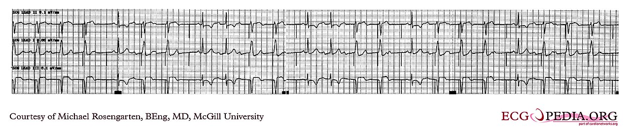
- Paced rhythm: wide QRS complexes can set off false alarms and get miscounted. Check the pulse
- Device guidance: follow the device clinic's and local protocol's electrode placement guidance
Post-Procedure Care & Transport
Immediate Aftercare
Once the leads are on, check the lead, the alarm limits and the baseline. If ST or QT was the reason for monitoring, record a 12-lead.
Ongoing Maintenance
- Electrodes: replace per protocol or when contact fails
- Skin: check under each electrode and move a damaged site
- Alarm limits: recheck after any change in rhythm, drug or condition
Transport Considerations
- Motion: movement and vehicle vibration create artifact. Recheck the leads after every move
- Alarm load: a moving patient and a busy vehicle mean more false alarms. Look at the patient first
- Drugs: when a QT-prolonging drug goes in during transport, recheck the QTc on a 12-lead
Document & Hand Off
- Why you monitored and which lead you used
- Alarm limits, and any change you made to them
- Baseline rhythm, ST and QTc, plus the time of the 12-lead
- Every alarm and event, and what you did about each
- Electrode sites and the condition of the skin
Clinical Pearls & Pitfalls
Pearls
- Confirm before acting: an ST or QT finding on the monitor needs a 12-lead
- Look at the patient: 88.8% of arrhythmia alarms were false in one ICU study. The real event is the one you can miss
- Switch leads deliberately: a small QRS in a lead means beats get undercounted
- Baseline first: an ST change needs a baseline from a stable tracing to be read against
Common Pitfalls
- Default alarm limits: individualize and document
- Trusting an ST trend after a lead change: reset the baseline first
- Assuming a quiet monitor means a normal QT: after a new drug, check the QTc on a 12-lead
- Silencing alarms at the bedside: pause, look at the patient, then respond
Education & Skill Maintenance
Skill Maintenance
- Practice lead placement on a partner and compare the strip with a 12-lead
- Practice setting an ST baseline on sample strips
- Practice recognizing a wide QRS, a small QRS and a noisy trace
- Debrief missed or delayed alarm responses using the unit's alarm log
References
- Sandau KE, Funk M, Auerbach A, et al. Update to Practice Standards for Electrocardiographic Monitoring in Hospital Settings: A Scientific Statement From the American Heart Association. Circulation. 2017;136(19):e273–e344. https://doi.org/10.1161/CIR.0000000000000527
- Drew BJ, Ackerman MJ, Funk M, et al. Prevention of torsade de pointes in hospital settings: a scientific statement from the American Heart Association and the American College of Cardiology Foundation. Circulation. 2010;121(8):1047–1060. https://doi.org/10.1161/CIRCULATIONAHA.109.192704
- Drew BJ, Harris P, Zègre-Hemsey JK, et al. Insights into the problem of alarm fatigue with physiologic monitor devices: a comprehensive observational study of consecutive intensive care unit patients. PLoS ONE. 2014;9(10):e110274. https://doi.org/10.1371/journal.pone.0110274
- American Association of Critical-Care Nurses. Ensuring Accurate ST-Segment Monitoring. Practice Alert. Published 2016; evidence update 2018. https://www.aacn.org/clinical-resources/practice-alerts/st-segment-monitoring
- Droperidol injection, solution. Prescribing information. American Regent, Inc. DailyMed; label effective 2023. https://dailymed.nlm.nih.gov/dailymed/drugInfo.cfm?setid=147e033d-d997-4ef6-8bb5-a9ba372590b2
- Morales-Arteaga JL, Meghdadi A, Wu D, et al. Alternative Lead ECG Placements. Ann Noninvasive Electrocardiol. 2026;31:e70176. https://doi.org/10.1111/anec.70176
- Macfarlane PW, Colaco R, Stevens K, Reay P, Beckett C, Aitchison T. Precordial electrode placement in women. Neth Heart J. 2003;11(3):118–122. https://pmc.ncbi.nlm.nih.gov/articles/PMC2499893/
- Celexa (citalopram hydrobromide) tablets. Prescribing information. Allergan, Inc. Revised 10/2023. DailyMed. https://dailymed.nlm.nih.gov/dailymed/drugInfo.cfm?setid=4259d9b1-de34-43a4-85a8-41dd214e9177
Medical Disclaimer
- For Educational Purposes Only: This content is intended for educational reference and should not be used for clinical decision-making.
- Not a Substitute for Professional Judgment: Always consult your local protocols, institutional guidelines, and supervising physicians.
- Verify Before Acting: Users are responsible for verifying information through authoritative sources before any clinical application.