

At a Glance
- Pattern
- Potassium, calcium and magnesium shifts that change conduction and repolarization. K⁺ sets the resting potential, Ca²⁺ the plateau, and low Mg²⁺ raises torsades risk.
- Normal (Adult)
- K⁺ 3.5–5.0 mEq/L, total Ca²⁺ 8.5–10.5 mg/dL, Mg²⁺ 1.7–2.2 mg/dL (lab-specific). ECG: T under 5 mm in limb leads, up to about 10 mm in precordial leads (shape matters more); no prominent U waves; QTc within sex limits.
- Confirm It
- T height against the QRS in the same lead (II, V2–V3); P-wave amplitude; QRS width; PR; QTc with separate U waves excluded. Confirm the level before acting on the pattern.
- Abnormal → Think
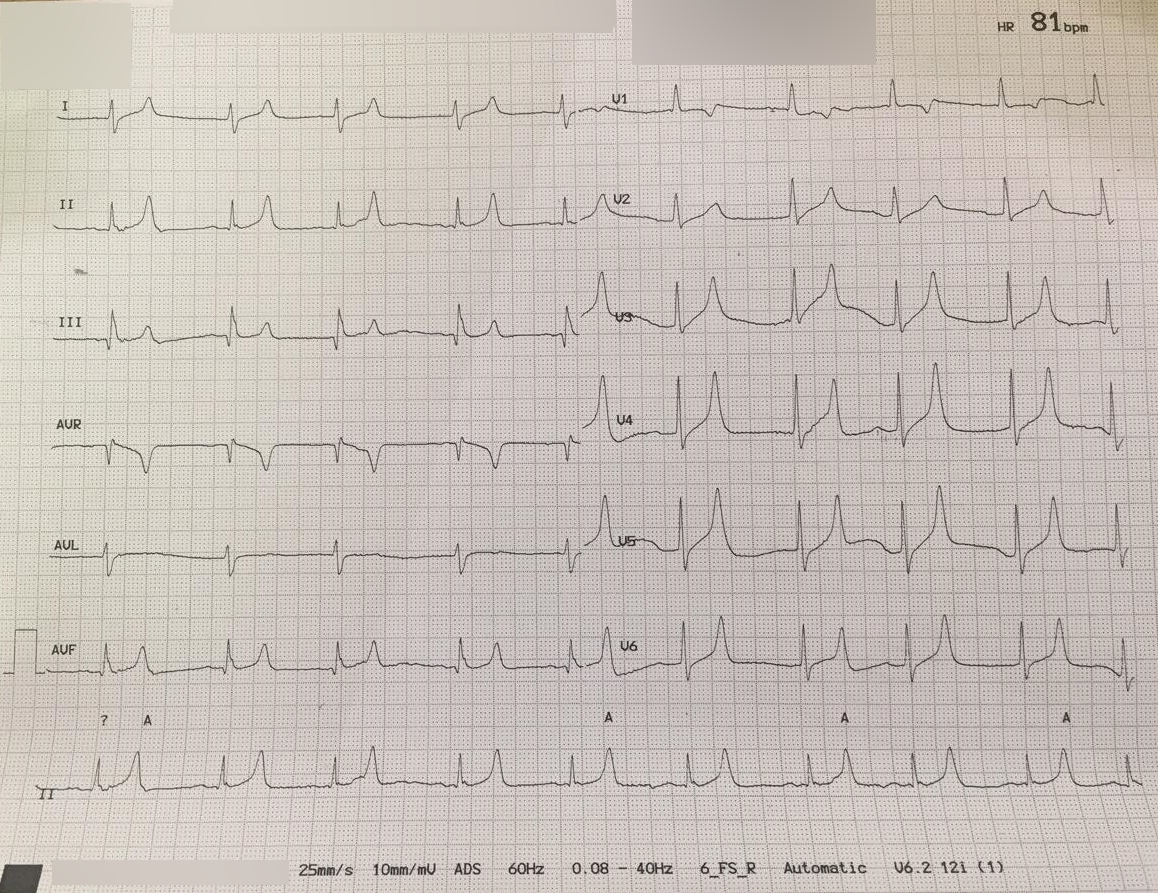
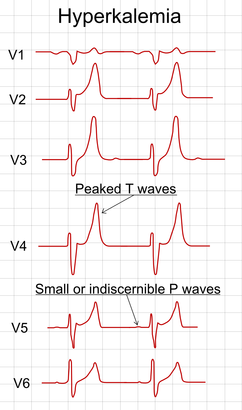
- Peaked T, flat P, long PR: hyperkalemia
- Wide QRS or sine wave: severe hyperkalemia
- Flat T, prominent U waves: hypokalemia
- Long QT with twisting VT: torsades (low K⁺, Mg²⁺ or drug)
- Red Flags
- Peaked T with wide QRS, PR prolongation, lost P waves or a sine wave: hyperkalemia emergency. Torsades: unsynchronized shock if pulseless or unstable; magnesium 2 g for recurrences.
- Bedside Action
- Hyperkalemic ECG: IV calcium now (gluconate by peripheral IV if pulse; chloride for arrest or central access), then insulin-dextrose; K⁺ on a non-hemolyzed sample. Torsades: shock if unstable; magnesium 2 g IV if recurrent; stop QT drugs.
Physiology & What It Represents
Every beat is ions moving across the myocardial cell membrane. Potassium sets the resting potential. Its movement out of the cell drives repolarization. When serum K⁺ climbs, the resting membrane sits partly depolarized. Sodium channels recover less, so conduction slows. Atrial muscle is more sensitive than ventricular muscle. The sinoatrial node and His bundle are the least sensitive. That is why atrial changes tend to show first.
Calcium comes in during the plateau of the ventricular action potential, and that drives contraction. It does not set the resting potential. So its main ECG footprint is the ST segment and the QT interval. Low magnesium is a well-established torsades risk. Low potassium and low magnesium both stretch repolarization unevenly. That unevenness is what sets up torsades de pointes.
Normal Values & How to Measure
| Parameter | Adult Normal | Pediatric Note | Where / How to Measure |
|---|---|---|---|
| Serum potassium (K⁺) | 3.5–5.0 mEq/L (3.5–5.0 mmol/L) | Use the age-specific lab range | Laboratory value on a non-hemolyzed sample; not an ECG measurement |
| Total calcium (Ca²⁺) | 8.5–10.5 mg/dL (2.12–2.62 mmol/L) | Use the lab's pediatric range | Ionized calcium is the better test when albumin is low |
| Magnesium (Mg²⁺) | 1.7–2.2 mg/dL (about 1.4–1.8 mEq/L) | Lab-specific | Serum level only; no ECG equivalent |
| T-wave amplitude | Under 5 mm in limb leads; up to about 10 mm in precordial leads (morphology matters more than height) | Juvenile T inversion in V1–V3 is normal | Tallest T against the QRS in the same lead (II, V2–V3) |
| QRS duration | ≤110 ms (≤0.11 s; about 2.75 small boxes) | Age-dependent | Widest lead; 120 ms or more is wide |
| PR interval | 120–200 ms (0.12–0.20 s; 3–5 small boxes) | Shorter in infants | P-wave onset to QRS onset |
| QTc | <450 ms men; <460 ms women | Age-specific limits | QRS onset to T-wave end, corrected; Fridericia or Framingham when HR is below 60 or above 90–100 |
- Pick the lead: compare each T wave with the QRS in the same lead, starting with II and V2–V3.
- Measure the T wave: amplitude from baseline in mm. A peaked T is tall, narrow-based and symmetric.
- Measure the QRS: width in the widest lead; 120 ms or more is wide.
- Measure the PR and check the P waves: P-wave onset to QRS onset, and whether every P has a QRS after it.
- Measure the QT: to the end of the T wave. Include a U wave only when it merges with the T wave.
Hyperkalemia (Peaked T to Sine Wave)
High potassium slows conduction and changes repolarization in a predictable order. That order is not always strict. The ECG does not measure the potassium level. It shows how far the heart has been pushed, and a tracing can change fast as K⁺ climbs. For the T-wave detail, see The T Wave.
ECG Criteria
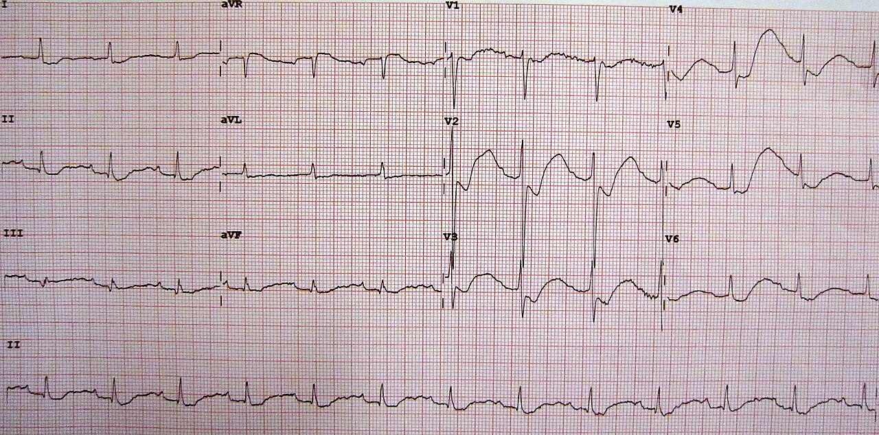
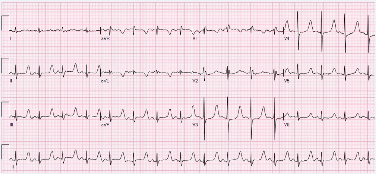
- Early: peaked T waves, tall and narrow-based with a pointed apex. Most common sign. Rarely life-threatening on its own.
- Atrial: P waves flatten, then disappear. The PR interval gets longer.
- Ventricular: the QRS widens. Wide QRS plus bradycardia is a late, high-risk pattern.
- Late: a sine-wave pattern. The QRS and T wave merge into one smooth undulating wave. Next comes ventricular fibrillation, asystole or PEA.
The changes may build over time. They do not always arrive in order, and several can show up at once.
Causes
- Kidney failure: the kidneys excrete less potassium. The most common setting.
- Medications: ACE inhibitors, ARBs, spironolactone and trimethoprim, alone or in combination.
- Shifts and load: acidosis, cell breakdown (crush injury, rhabdomyolysis), hemolysis and potassium-rich fluids or supplements.
Clinical Significance
- Insensitive: one retrospective hospital series found the ECG insensitive for hyperkalemia. No T-wave threshold reliably split patients with it from those without.
- Severity: a 2026 UK guideline reports ECG features in about 66% of patients with K⁺ of 6.5 mEq/L or higher. A normal ECG can still come with severe hyperkalemia.
- Sine wave: a sine wave with muscle weakness or bradycardia is time-critical. Get the potassium now and start treatment.
Hypokalemia (Flat T and Prominent U Waves)
Look for a small deflection after the T wave, most visible in V2–V3. That is the U wave, and it gets prominent when potassium drops. Low potassium makes repolarization uneven. Hypokalemia is one of the best-established risk factors for torsades de pointes.
ECG Criteria
- T waves: they flatten first, then invert in several leads. ST depression often comes with them.
- U waves: they get prominent. They can merge with the T wave into a camel-hump shape that looks like a long QT.
- PR and rhythm: the PR may lengthen slightly. Ectopy and arrhythmia get more likely as the level falls.
Causes
- Diuretics: loop and thiazide diuretics are the common cause in hospital and at home.
- GI losses: vomiting, nasogastric suction and diarrhea.
- Shifts: insulin, beta-agonists such as albuterol, and alkalosis.
- Low magnesium: often comes with it. Check and correct both. Low magnesium is a torsades risk factor on its own.
Clinical Significance
- Prominent U waves with a long QT: check K⁺ and Mg²⁺ now, before any QT-prolonging drug is given.
- Replacement: per local protocol, with continuous ECG monitoring. Concentrated IV potassium is high-alert. It goes by pump only.
Hypocalcemia and Hypercalcemia (Calcium and the QT)
Calcium carries the plateau of the ventricular action potential. Low calcium lengthens the ST segment and QT. High calcium shortens them. The QT effect is real. Torsades from low calcium alone is rare.
ECG Criteria
- Hypocalcemia: a long ST segment with a relatively normal T wave, and a prolonged QT.
- Hypercalcemia: a shortened ST segment and a short QT (see QT Interval).
Causes
- Low calcium: low albumin (the total is misleading), citrate after transfusion, sepsis or pancreatitis, low Mg²⁺, and loss of parathyroid function after thyroid surgery.
- High calcium: malignancy, primary hyperparathyroidism, vitamin D excess, thiazide diuretics and lithium.
Clinical Significance
- Measure ionized calcium when albumin is low. Total calcium can misclassify the state.
- Calcium is not a potassium treatment: IV calcium given for hyperkalemia protects the myocardium. It does not lower K⁺.
Hypomagnesemia and Torsades de Pointes
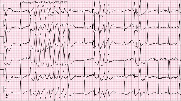
In torsades de pointes, the QRS axis twists around the baseline in bursts. It is polymorphic VT riding on a long QT. The main reversible triggers: low magnesium, low potassium and QT-prolonging drugs.
ECG Criteria
- Baseline QT: a QTc of 500 ms or more, or a rise of 60 ms or more from baseline, puts torsades risk high.
- Rhythm: bursts of polymorphic VT, often after a pause or a short-long-short run (see Ventricular Rhythms).
- Hypomagnesemia on the ECG: not specific. It can look just like hypokalemia, with flat T waves, U waves and a long QT.
Causes
- Losses: loop and thiazide diuretics, alcohol, diarrhea and GI losses.
- Drugs that waste magnesium: aminoglycosides, amphotericin B, cisplatin and cyclosporine.
Clinical Significance
- Recurrent torsades: IV magnesium for recurrences with a long QT (AHA 2025 Part 9, COR 2b, C-LD). AHA 2025 gives no dose, rate or serum-level condition. Dose used on this page: 2 g IV, diluted, over 10 minutes with a pulse (NASEMSO v3.0, Tachycardia with a Pulse, p. 44: 1–2 g over 10 minutes; that source gives no repeat); further doses per medical direction. Pulseless: 2 g IV/IO over 1–2 minutes (NASEMSO v3.0, p. 119).
- Remove the cause: stop QT-prolonging drugs and correct K⁺ and Mg²⁺. Pause-dependent torsades may need overdrive pacing.
- Pulseless or unstable: unsynchronized defibrillation first (AHA 2025 Part 9, COR 1). Magnesium comes second. Do not delay the shock to give it.
Differential Diagnosis & Mimics
| Feature | Hyperkalemia | Hyperacute T (Occlusion MI) | Hypokalemia |
|---|---|---|---|
| T wave | Tall, narrow-based, symmetric, pointed | Tall, broad T in one coronary territory | Flat, then inverted |
| Distribution | Diffuse, across territories | One territory, with reciprocal change | Diffuse, anterior leads most visible |
| P wave and PR | Flattened; PR prolonged | Normal | Normal, PR may lengthen slightly |
| QRS | Widens late, then sine wave | Normal | Normal |
| Setting | Kidney failure, potassium-raising drugs, acidosis | Chest pain and ACS risk factors | Diuretics, vomiting, diarrhea |
Artifact & Lead Misplacement
- Limb-lead reversal can invert or flatten leads and mimic a T-wave problem.
- Precordial electrode placement high or low changes the T and U waves in V2–V3.
- Shivering, vibration and movement in a moving vehicle can mimic peaked or flattened T waves.
- Paced rhythms change the ST and T waves. Check the pacing spike and capture before you read ischemia.
Normal Variants
- Tall, symmetric T waves can be physiologic in some athletes, and in people with no disease at all. Judge by morphology, prior ECGs and the potassium level.
Bedside Management & Escalation
Your local protocols and medical direction govern treatment. The adult non-arrest doses here are from the 2026 UK Kidney Association guideline (UKKA). The arrest insulin-dextrose dose is from the 2025 AHA Guidelines for CPR and ECC, Part 10. Pediatric calcium chloride and magnesium doses are from NASEMSO v3.0 (see the dosage table). Every other pediatric dose is weight-based and protocol-dependent, so confirm it against the current PALS reference or local protocol.
| Situation | BLS (EMT) | ALS (AEMT / Paramedic / RN) | Critical Care / Transport |
|---|---|---|---|
| Suspected hyperkalemia with ECG changes, pulse present | 12-lead and monitor where allowed; oxygen if SpO₂ <94%; ask about kidney failure and potassium-raising drugs; notify early | IV calcium now for ECG changes (calcium gluconate 30 mL of 10% over 10 min by peripheral IV; calcium chloride 10 mL over 5 min if peri-arrest or with central or deep access); obtain K⁺ (point-of-care blood gas if available); insulin 10 units with dextrose 25 g | Continuous monitoring (3-lead minimum); serial ECG; urgent dialysis or potassium removal; early notice to critical care |
| Bradycardia or AV block with hyperkalemia | Pads on for transport; oxygen; early ALS | Calcium first; transcutaneous pacing per protocol if the rate is unstable | Check capture by pulse and SpO₂ waveform; pacing per Transcutaneous Pacing |
| Pulseless, hyperkalemia suspected | CPR and defibrillation for VF per BLS | ALS arrest algorithm; calcium chloride 10 mL (1 g) of 10% IV/IO push (UKKA 25.1; AHA 2025 Part 10, COR 2b); insulin 10 units IV over 15–30 min with D50 50 g (AHA 2025 Part 10, Table 3, COR 2b; UKKA and ERC use 25 g); sodium bicarbonate 50 mEq IV/IO (UKKA 25.3; AHA 2025 Part 10, COR 2b); no albuterol (AHA 2025 Part 10, COR 3) | Dialysis during CPR may be considered if medical therapy fails and staff are available |
| Torsades with a pulse | Recognize; pads on; oxygen; notify | Magnesium 2 g IV/IO diluted over 10 min for recurrent torsades with long QT (NASEMSO v3.0, Tachycardia with a Pulse, p. 44: 1–2 g over 10 min; AHA 2025 Part 9, COR 2b, gives no dose or rate); further doses per medical direction; stop QT drugs; unsynchronized shock if unstable (COR 1 for sustained polymorphic VT) | Overdrive pacing for pause-dependent torsades; correct K⁺ and Mg²⁺ |
| Pulseless torsades | CPR and defibrillation without delay | Unsynchronized defibrillation without delay (AHA 2025 Part 9, COR 1); magnesium 2 g IV/IO over 1–2 min (NASEMSO v3.0, p. 119). AHA 2025 advises against routine magnesium in adult arrest (COR 3), so never delay a shock for it | Treat the cause; hand off every QT-prolonging drug |
| Hypokalemia or hypomagnesemia, no arrhythmia | 12-lead if allowed; monitor | Check K⁺ and Mg²⁺; replace per protocol | Continuous ECG; repeat QTc after replacement; hold QT-prolonging drugs |
| Therapy | Adult | Pediatric | Key Notes |
|---|---|---|---|
| Calcium gluconate 10% (Calcium Gluconate) | 30 mL (3 g) IV/IO slowly over about 10 min for ECG changes with a pulse; repeat if changes persist Same calcium as 10 mL of calcium chloride (6.8 mmol). Repeat the ECG after each dose; effect lasts about 60 minutes. Differs from FDA label: the US labels list only hypocalcemia and cap the rate (calcium gluconate 200 mg/min for adults, so 3 g takes at least 15 min; calcium chloride 1 mL/min as a slow infusion, central or deep vein). The hyperkalemia rate here (gluconate 30 mL over 10 min, chloride 10 mL over 5 min) is from UKKA 2026 and is off-label. |
Protocol-dependent; weight-based (see Weight-Based Dosing) Confirm against the current PALS reference or local protocol |
Preferred calcium salt for a patient with a pulse and ECG changes (UKKA 16.2b), and the usual choice for a peripheral IV. Protects the myocardium; does not lower K⁺. Start K⁺-shifting therapy at the same time. |
| Calcium chloride 10% (Calcium Chloride) | 1 g (10 mL of 10%) IV/IO for ECG changes in cardiac arrest or peri-arrest, or with central or deep access (UKKA 16.2b); repeat if changes persist 10 mL (1 g) over 5 min (peri-arrest or central/deep access; UKKA); label limit is 1 mL/min (differs from FDA label). |
20 mg/kg (0.2 mL/kg of 10%) IV/IO over 5 min Max 1 g per dose. Source: NASEMSO v3.0, p. 119 (20 mg/kg); rate and 1 g maximum from NASEMSO p. 82 (adult values, no faster than 1 mL/min) |
Preferred salt in arrest. Label: give into a central or deep vein; do not give IM or SC. Extravasation can cause tissue necrosis, so avoid a peripheral infusion when another salt is suitable and check the line. Protects the myocardium; does not lower K⁺. Label: avoid with digoxin; if unavoidable, monitor ECG and calcium closely. Start K⁺-shifting therapy at the same time. |
| Regular insulin with dextrose (Insulin; Dextrose) | 10 units IV with 25 g dextrose for K⁺ 6.5 mEq/L or higher (suggested at 6.0–6.4) Consider a second dose if K⁺ is not below 6.0 within 2 hr (UKKA p.98); each repeat adds hypoglycemia risk. If pre-treatment glucose is under 7.0 mmol/L (126 mg/dL), add D10 at 50 mL/hr for 5 hours |
0.1 units/kg IV over 15–30 min with D10 5 mL/kg (0.5 g/kg) IV bolus, stated for cardiac arrest (see Weight-Based Dosing); non-arrest use per pediatric protocol Maximum 10 units for severe hyperkalemia (AHA 2025 Part 10, Table 3) |
High-alert Moves K⁺ into cells; it does not remove it. Check glucose at 0, 30, 60, 90, 120, 180, 240, 300 and 360 min (UK schedule). Hypoglycemia risk is higher in kidney failure and low body weight. Some US protocols use 5 units or 0.1 units/kg. A pooled analysis of 10 retrospective studies found less hypoglycemia with lower doses and no difference in K⁺ fall; a later single-center study found 10 units lowered K⁺ more (0.9 vs 0.63 mmol/L); UKKA 2026 states more study is needed before 5 units can be recommended. This site uses 10 units with the glucose rules above. |
| Albuterol, nebulized (Albuterol) | 10–20 mg nebulized as an adjunct for K⁺ 6.5 mEq/L or higher (UK guidance) Adjunct only; not monotherapy for severe hyperkalemia |
Per local pediatric hyperkalemia protocol; not the asthma dose on the Albuterol page |
Shifts K⁺ as an add-on to calcium and insulin, never alone. Not used in cardiac arrest (AHA 2025 Part 10, COR 3). Differs from FDA label: off-label dose (label nebulized dose is 2.5 mg for asthma). 10–20 mg is from UKKA 2026. The E3 Albuterol page gives asthma dosing only. |
| Sodium bicarbonate (Sodium Bicarbonate) | 50 mEq (one 50 mL vial) IV/IO when acidemic or per arrest protocol Not used routinely for hyperkalemia alone (UK guidance); repeat per blood gas and protocol |
1 mEq/kg IV/IO over 5 min Max 50 mEq per dose. Source: NASEMSO v3.0, p. 82 (1 mEq/kg, max 50 mEq, IV bolus over 5 min) |
Shifts K⁺ into cells when acidemic; removes none. Flush the line between calcium and bicarbonate; they precipitate if mixed. |
| Magnesium sulfate (Magnesium Sulfate) | 2 g IV/IO, diluted, over 10 min for recurrent torsades with a pulse and long QT (NASEMSO v3.0, p. 44: 1–2 g over 10 min); further doses per medical direction No fixed max; check reflexes, respiratory rate and serum Mg²⁺ after repeat doses. Pulseless: 2 g over 1–2 min (NASEMSO v3.0, p. 119) |
25–50 mg/kg IV/IO for torsades (NASEMSO v3.0, p. 119) Max 2 g per dose. Hypomagnesemia without torsades: per pediatric protocol |
High-alert For recurrent torsades with a long QT (AHA 2025 Part 9, COR 2b, C-LD); routine magnesium in adult arrest is not recommended (COR 3). Stop QT-prolonging drugs and correct K⁺. Give in grams and mL aloud; the 50% vial is 500 mg/mL. |
| Potassium replacement (hypokalemia) | Oral or IV per protocol, with ECG and K⁺ monitoring |
Per pediatric protocol; weight-based |
High-alert Concentrated IV potassium must be diluted and given by pump only, never as an IV push or undiluted. Rate limits are in the product label and protocol. |
Local protocols and online medical direction come first.
Pediatric & Special Populations
Pediatrics
- Weight-based doses: calcium, insulin, bicarbonate and magnesium all run on weight. Check the per-dose maximum on Weight-Based Dosing.
- Normal values differ: rates, PR and T-wave shape shift with age. Juvenile T inversion in V1–V3 is normal through adolescence. Set it against prior ECGs and the age norms on Vital Signs.
- Hyperkalemia in children: same ECG sequence. Treat by pediatric protocol and recheck the potassium early.
Other Populations
- Kidney failure and dialysis: with severe hyperkalemia and toxic ECG changes, give IV calcium even when dialysis is right there. Dialysis itself is urgent (see Hemodialysis).
- Pacemakers and ICDs: hyperkalemia widens the paced QRS and raises thresholds. That can cause failure to capture, particularly above about 7.0 mEq/L. A paced rhythm does not protect against hyperkalemia.
- Older adults: diuretic-related hypokalemia and hypomagnesemia are common. Older patients also often take more QT-prolonging drugs.
- Heart disease and QT-prolonging drugs: underlying heart disease and several QT-prolonging drugs raise torsades risk. The same electrolyte shift matters more in those patients (see QT Interval).
Monitoring & Transport Considerations
| Issue | What to Do |
|---|---|
| Monitoring | Continuous 3-lead monitoring at minimum for K⁺ of 6.5 mEq/L or higher (UKKA 14.2, 26.2), for any ECG feature of hyperkalemia on the 12-lead, and for K⁺ 6.0–6.4 mEq/L when the patient is clinically unwell or a rapid rise is expected (UKKA 14.2) |
| Monitor lead | Lead II for P waves; V2–V3 for T and U waves; the lead with the clearest T wave for QT trending |
| 12-lead | Repeat after IV calcium, after each potassium-shifting drug, and with any rhythm change |
| Potassium trend | Serum K⁺ at about 1, 2, 4, 6 and 24 h after treatment, because rebound hyperkalemia occurs as the shift wears off (UK guidance) |
| Glucose after insulin | Check at 0, 30, 60, 90, 120, 180, 240, 300 and 360 min (UK schedule) |
| Pacing | Check capture by pulse and SpO₂ waveform; hyperkalemia raises pacing thresholds |
| Blood sample | Non-hemolyzed sample; hemolysis and pseudohyperkalemia falsely raise K⁺. Use the lab sample for the decision and a point-of-care gas while waiting, if available |
| Transport and handoff | Record the time of each calcium dose, insulin and dextrose dose, and each potassium result. Notify the receiving team early |
Step-by-Step Approach & Pitfalls
- Get a 12-lead ECG as early as allowed, and compare each T wave with the QRS in the same lead.
- Measure the QRS and PR, and look for flat or absent P waves.
- Check the rhythm for a sine wave, bradycardia, AV block or a VT pattern.
- If the QT looks long, separate any U waves before measuring, and draw K⁺, Mg²⁺ and Ca²⁺ on a non-hemolyzed sample.
- Ask about causes: kidney failure, ACE inhibitors, ARBs, spironolactone, diuretics, vomiting and diarrhea.
- For hyperkalemic ECG changes, give IV calcium first, then insulin 10 units with dextrose 25 g (see Bedside Management), and repeat the ECG.
- For torsades, unsynchronized shock if pulseless or unstable, magnesium 2 g IV for recurrences, and removal of QT-prolonging drugs.
Common Pitfalls
- A normal-looking ECG does not prove the potassium is normal.
- Waiting for the potassium result before calcium, when the ECG already shows hyperkalemic change.
- Calling a peaked T wave ischemia before anyone checks the potassium.
- Giving amiodarone or procainamide for torsades. Both lengthen the QT.
- Calling a fused U wave a long QT before checking the electrolytes.
- Correcting potassium and forgetting magnesium. Torsades can persist.
ECG Examples
Clinical Pearls
References
- Cao D, Arens AM, Chow SL, et al. Part 10: Adult and pediatric special circumstances of resuscitation: 2025 American Heart Association guidelines for cardiopulmonary resuscitation and emergency cardiovascular care. Circulation. 2025;152(16 suppl 2):S578–S672. https://doi.org/10.1161/CIR.0000000000001380
- Lasa JJ, Dhillon GS, Duff JP, et al. Part 8: Pediatric advanced life support: 2025 American Heart Association and American Academy of Pediatrics guidelines for cardiopulmonary resuscitation and emergency cardiovascular care. Circulation. 2025;152(16 suppl 2):S479–S537. https://doi.org/10.1161/CIR.0000000000001368
- UK Kidney Association. Treatment of acute hyperkalaemia in adults: clinical practice guideline. Updated July 2026. https://www.ukkidney.org/sites/default/files/documents/FINAL%20VERSION%20-%20UKKA%20CLINICAL%20PRACTICE%20GUIDELINE%20-%20MANAGEMENT%20OF%20HYPERKALAEMIA%20IN%20ADULTS%20-%20UPDATED%20JULY%202026_0.pdf
- National Association of State EMS Officials (NASEMSO). National Model EMS Clinical Guidelines, Version 3.0. March 2022. General Medical, Hyperglycemia (p. 82, hyperkalemia calcium); Cardiovascular, Tachycardia with a Pulse (p. 44, torsades magnesium 1–2 g over 10 min); Resuscitation, Cardiac Arrest (p. 119, torsades magnesium and reversible-cause calcium doses). Page numbers are PDF pages (printed page numbers are one lower). Accessed October 15, 2026. https://hhs.iowa.gov/media/198/download
- Montague BT, Ouellette JR, Buller GK. Retrospective review of the frequency of ECG changes in hyperkalemia. Clin J Am Soc Nephrol. 2008;3(2):324–330. https://doi.org/10.2215/CJN.04611007
- Hüser C, Terporten J, Macherey-Meyer S. ECG in hyperkalemia. Dtsch Arztebl Int. 2025;122(17):466. https://doi.org/10.3238/arztebl.m2025.0117
- Hospira, Inc. Calcium chloride injection, USP 10%: prescribing information. DailyMed, U.S. National Library of Medicine; revised January 2026. https://dailymed.nlm.nih.gov/dailymed/drugInfo.cfm?setid=c4c65e48-85f8-4dcf-6281-06e40959cc79
- Somers MP, et al. The prominent T wave: electrocardiographic differential diagnosis. Am J Emerg Med. 2002;20(3):243–251. PMID 11992348. https://pubmed.ncbi.nlm.nih.gov/11992348/
- 2025 American Heart Association Guidelines for CPR and ECC. Part 9: Adult Advanced Life Support. Recommendations for polymorphic VT and torsades (COR 1 for immediate unsynchronized shock; COR 2b for magnesium in recurrences with long QT; COR 3 for routine magnesium in arrest). Accessed October 15, 2026. https://cpr.heart.org/en/resuscitation-science/cpr-and-ecc-guidelines/adult-advanced-life-support
- Moussavi K, Garcia J, Tellez-Corrales E, Fitter S. Reduced alternative insulin dosing in hyperkalemia: a meta-analysis of effects on hypoglycemia and potassium reduction. Pharmacotherapy. 2021;41:598–607. https://doi.org/10.1002/phar.2596
- Finder SN, McLaughlin LB, Dillon RC. 5 versus 10 units of intravenous insulin for hyperkalemia in patients with moderate renal dysfunction. J Emerg Med. 2022;62:298–305. https://doi.org/10.1016/j.jemermed.2021.10.027
- Fresenius Kabi USA. Calcium gluconate injection: prescribing information. Revised 6/2023 (current DailyMed version, checked October 15, 2026). DailyMed, U.S. National Library of Medicine. https://dailymed.nlm.nih.gov/dailymed/drugInfo.cfm?setid=8b77c3d2-992d-4261-8421-9cfd07328fbf
Medical Disclaimer
- For Educational Purposes Only: This content is intended for educational reference and should not be used for clinical decision-making.
- Not a Substitute for Professional Judgment: Always consult your local protocols, institutional guidelines, and supervising physicians.
- Verify Before Acting: Users are responsible for verifying information through authoritative sources before any clinical application.